

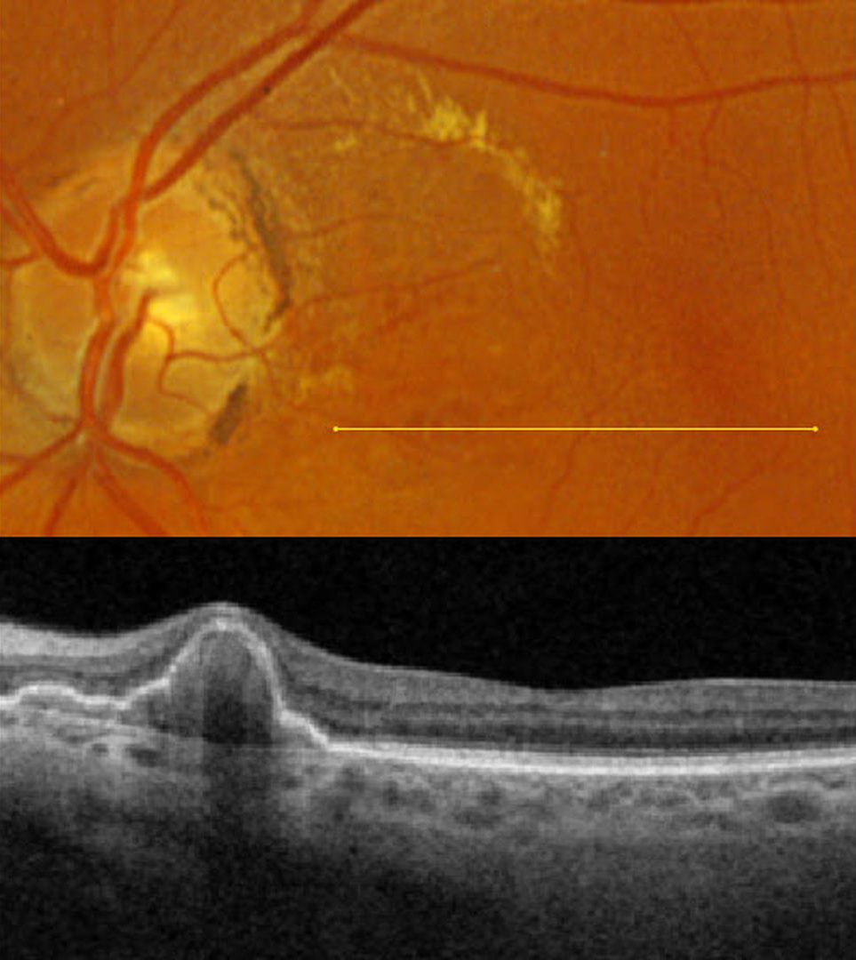
This 67YO female presented for a second opinion regarding possible age-related macular degeneration (AMD). She was without acute visual complaints. Vision was 20/25 in her normal OD and 20/30 OS.
Optos color RGB imaging shows macula lipid in the superonasal macula. Several red subretinal nodules are present inferior to the lipid. Triton swept-source OCT through these lesions shows a nodular and somewhat multi-lobulated RPE detachment (PED). We will follow her closely for any increase in exudation.
Learning Points:
At the Macula Society meeting in 1983 and at the Academy of Ophthalmology meeting in 1984, Brucker et al presented 7 patients with multiple recurrent sub-RPE and subretinal hemorrhages, which they termed the posterior uveal bleeding syndrome. This was followed by multiple authors reporting similar findings (Stern et al AJO 1985;100:560-569, Kleiner et al Retina 1990;10:9-17, and Yannuzzi et al Retina 1990;10:1-8). Dr. Gass, who was assigned as the reviewer for Yannuzzi’s manuscript titled “Idiopathic Polypoidal Choroidal Vasculopathy,” took issue with calling these lesions ‘polyps’ (Alexander J Brucker, personal communication 2/12/26), which are defined by Stedman’s Medical Dictionary as growths that protrude from a mucous membrane. When Yannuzzi challenged Dr. Gass for a better term, he could not come up with one; Yannuzzi’s terminology won out, and the rest is history. In our patient’s case, the clinical red ‘polyps’ corresponded to peaked, multilobulated RPE detachments on OCT.
PCV is likely distinct from age-related macular degeneration, occurring in a different population (more common in African Americans and Asians), with a relative lack of drusen and a thicker choroid. In our experience, many patients with ‘wet AMD’ actually have PCV or pachychoroid neovascularization.



