

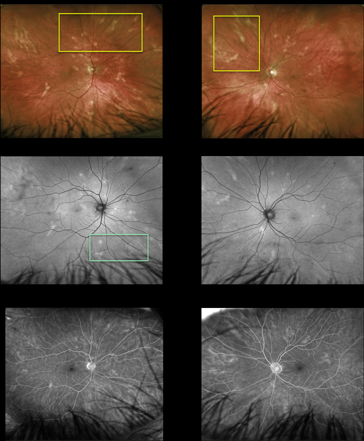
This healthy 51YO male presented with asymptomatic lesions in each fundus. Vision was 20/25 OU.
Multimodal imaging findings were symmetrical bilaterally. Optos color imaging shows amelanotic subretinal lesions that seem to radiate outwards from each disc. These lesions are mostly hyperautofluorescent with window-type defects on fluorescein angiography. OCT scanning was normal bilaterally (not shown). HLA-A29 was negative.
Learning Points:
Birdshot chorioretinopathy was appropriately named and described by Drs. Ryan and Maumenee (AJO 1980;89:31-45), since the lesions look like shotgun birdshot scattered throughout the fundus. Birdshot is typically chronic and bilateral, with a very high HLA-A29 association.
We are not certain that our patient has birdshot. The pattern of lesions and less creamy coloration is somewhat atypical of what we’ve normally seen in our practice.
Although HLA-A29 is positive in about 90% of cases, it is not required for the diagnosis in the setting of other clinical findings (SUN Working Group AJO 2021;228:65-71). Finally, birdshot lesions are typically hypofluorescent (Koizumi et al., Ophthalmology 2008;115:e15-e20), unlike the hyperfluorescence observed in our patient.
Since he is asymptomatic, has no signs of active uveitis, and no macular edema, we will initially follow him twice yearly.



