

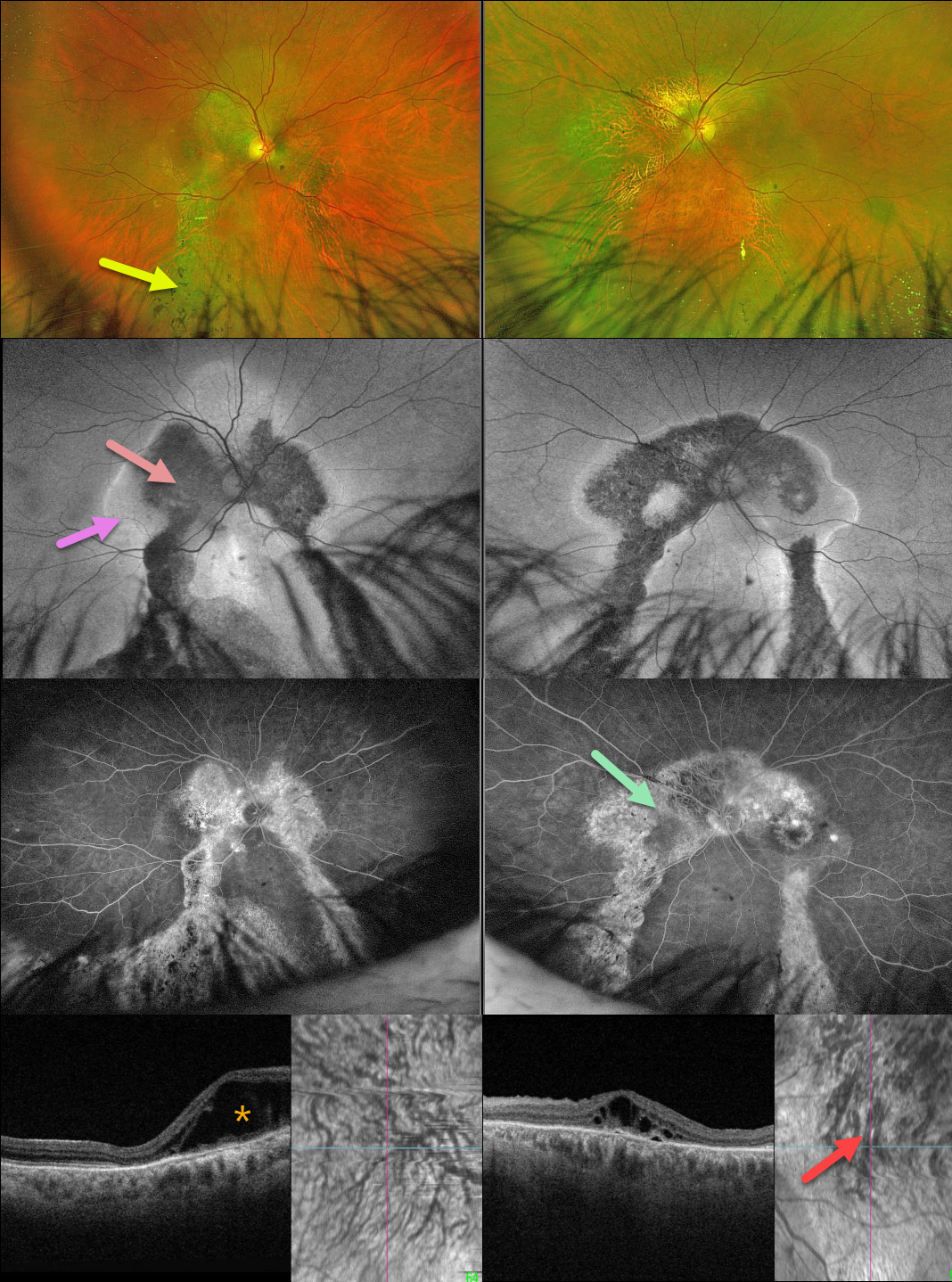
This healthy 62YO male presented with a several-year history of bilateral vision loss. There was no family history of eye disease. Vision was 20/400 OU.
Optos color RG imaging shows bilateral, symmetrical peripapillary and macular pigmentary changes extending into the gutters towards the inferior peripheries. Intraretinal pigment migration (spicules) is noted inferiorly.
On fundus autofluorescence (FAF), these areas are hypo-FAF centrally with hyper-FAF along their margins, and exhibit window defects on fluorescein angiography.
Outer retinal fluid is noted nasally OD and centrally OS on Triton swept-source OCT. En face OCT shows bilateral choroidal pachyvessels with anastomoses. Genetic testing was completely negative.
Learning Points:
Gutters are commonly seen in pathology with chronic subretinal leakage, including central serous retinopathy, choroidal hemangioma, choroidal nevus, and uveal melanoma. These pigmentary changes are usually best imaged with FAF. Our patient’s OCT certainly shows pachychoroid features, but the symmetrical double gutters in each eye would be quite unusual for central serous retinopathy.
Our patient’s gutters are somewhat reminiscent of a patient we have previously posted with the Senior-Løken Syndrome, although this is associated with renal failure and an NPHP1 mutation, which our patient did not have.



