

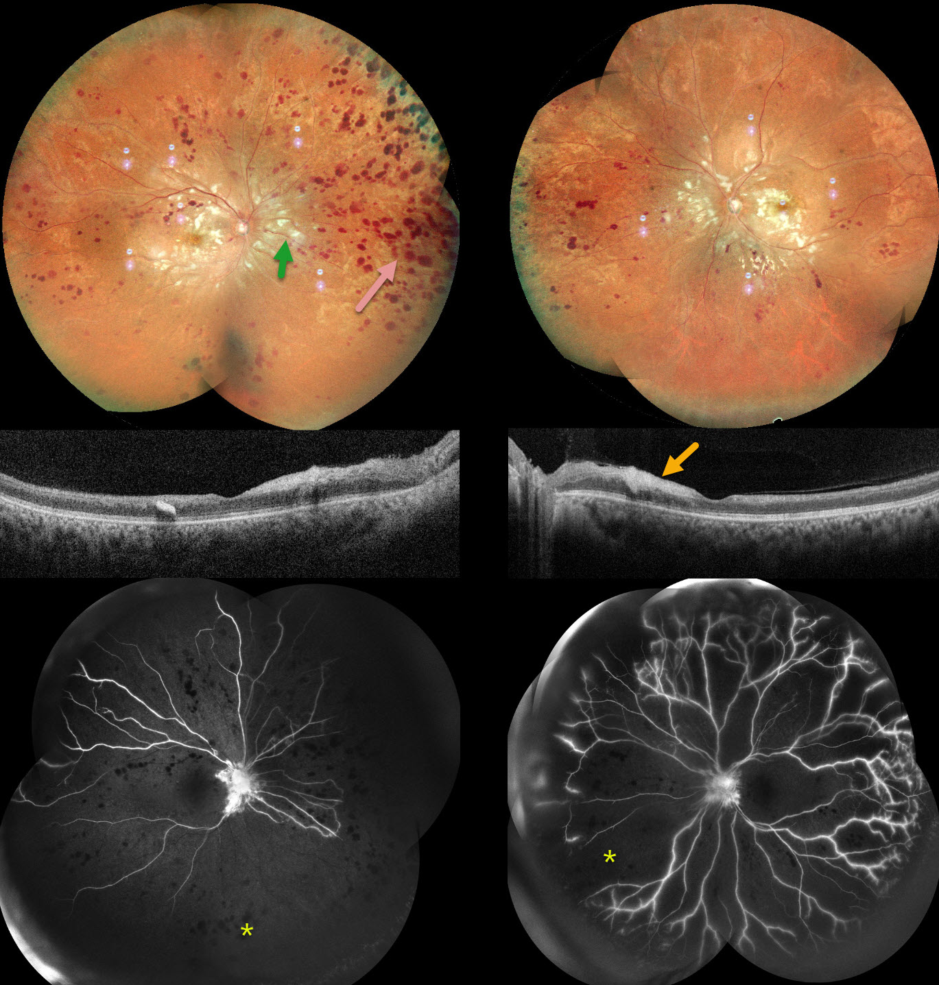
This previously healthy 32YO male presented with a 3-month history of bilateral vision loss following a viral illness. He was treated elsewhere for post-fever retinitis with intravitreal triamcinolone and oral steroids with antivirals. He also had a right lobe pneumonia. Testing was positive for CMV IgG and IgM and negative for TB.
When examined in our office, vision was 20/200 OU. Anterior segments were normal, with mild bilateral vitreous cells.
Pseudocolor SLO imaging shows extensive cotton-wool spots throughout each posterior pole, with more distal retinal hemorrhages that extend into the peripheries. OCT scanning shows possible vitreous cells with variable inner retinal thickened/thinned hyperreflectivity. Fluorescein angiography shows profound bilateral ischemia and disc leakage. We advised bilateral panretinal photocoagulation, but he was unfortunately immediately lost to follow-up.
Learning Points:
Epidemic (post-fever) retinitis is an infectious or para-infectious entity that develops in immunocompetent individuals several weeks following a bacterial, viral, or protozoal infection. Causes include Dengue, Rickettsiosis, West Nile, Leptospirosis, Chikungunya, Typhoid, and Ebola (none of which were tested in our patient).
Patients present with sudden, painless vision loss. Fundus findings include unilateral or bilateral unifocal or multifocal patches of retinitis with possible nerve involvement, serous macular detachment, and vasculitis or frosted branch angiitis. There is usually a favorable response to steroids and treating the underlying infectious cause with appropriate systemic therapy. See Mahendradas et al (Indian J Ophthalmol 2020;68:1775-1786) for an excellent post-fever retinitis review.



