

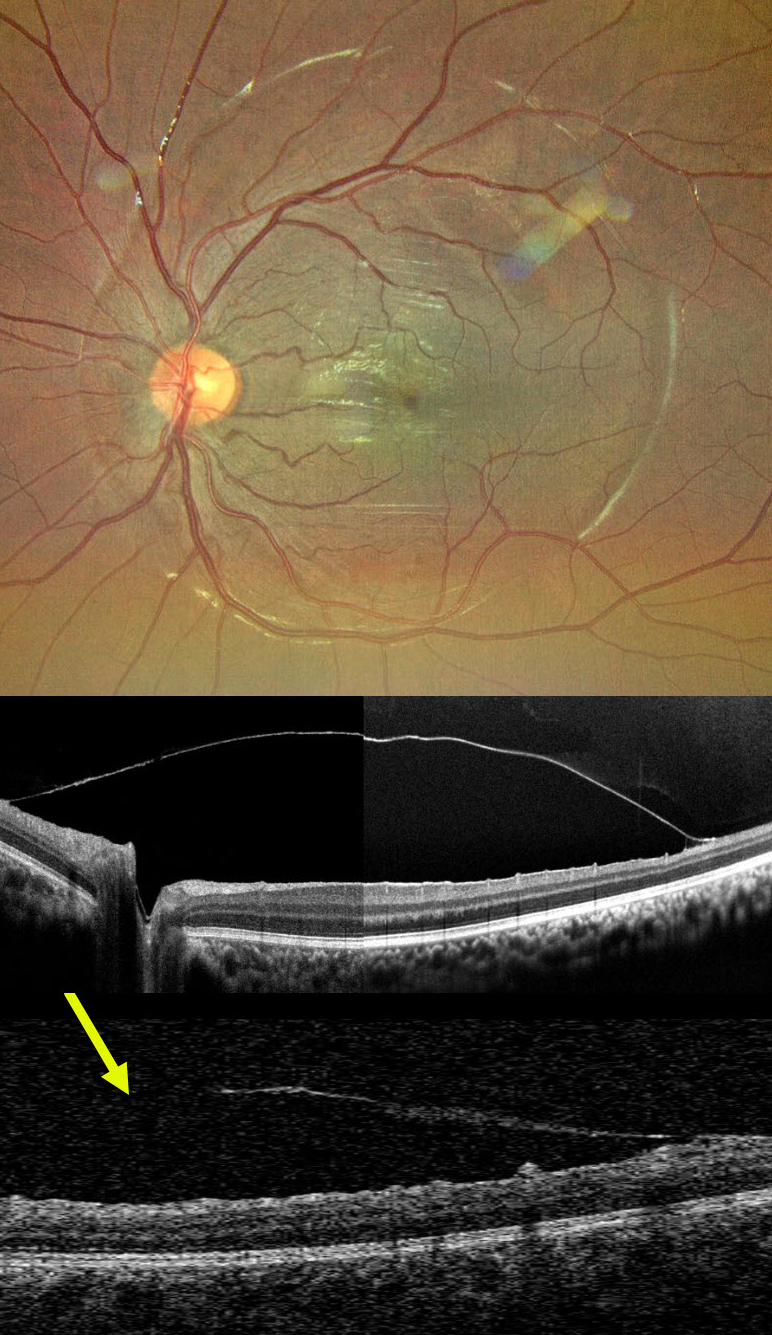
This 12YO boy came in for a routine eye examination. Although vision was 20/20 bilaterally, the right fundus showed a striking premacular sheen.
Ultra-widefield imaging reveals a round internal limiting membrane (ILM) elevation surrounding the posterior pole. On further questioning, he gave a history of blunt trauma to the right eye 2 years earlier that was treated elsewhere with the ND:YAG laser. The opening in the ILM is noticed inferiorly on OCT.
This case demonstrates a presumed chronic and stable sub-ILM detachment likely secondary to prior sub-ILM hemorrhage. Fortunately, he remains completely asymptomatic with perfect vision and no distortion.
A valuable reminder that retinal imaging can unearth forgotten chapters of ocular history, and that fundus examination with multimodal imaging can reveal prior stories.



