

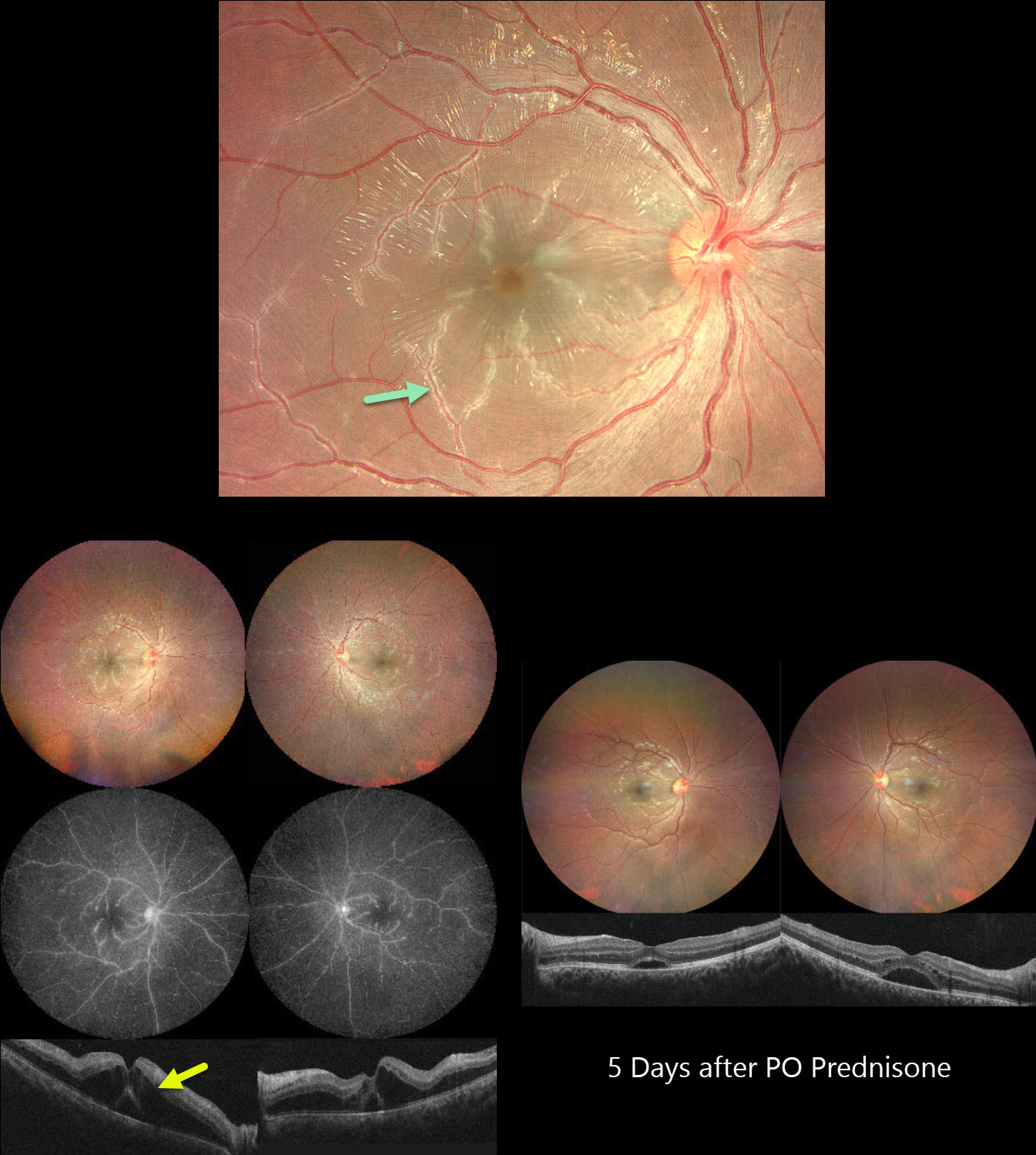
This previously healthy 21YO male developed bilateral vision loss 1 week following a viral illness. Vision was 20/100 OU.
Color photography shows a bilateral frosted-branch appearance of the retinal veins, most prominent in the maculas. Fluorescein angiography shows diffuse leakage and staining of the venous vascular tree. OCT scanning shows marked bilateral outer nuclear layer edema with foveal subretinal fluid. He was started on oral prednisone 60mg daily. Five days later vision improved to 20/40 OU. The venous changes are dramatically better with decreasing macular fluid. A slow steroid taper began.
Learning Points:
Frosted branch angiitis is a retinal vasculitis that gets its name from the involved vessels resembling branches of a tree that are “frosted” with snow (Kleiner et al, AJO 1988;106:27-34). Although these findings may be isolated, they are often considered a phenotype found in many inflammatory conditions, most commonly CMV retinitis. The inflammation is usually highly responsive to steroids, and the visual prognosis is generally good, although some may develop secondary neovascularization in the anterior and posterior segments due to widespread ischemia.



