

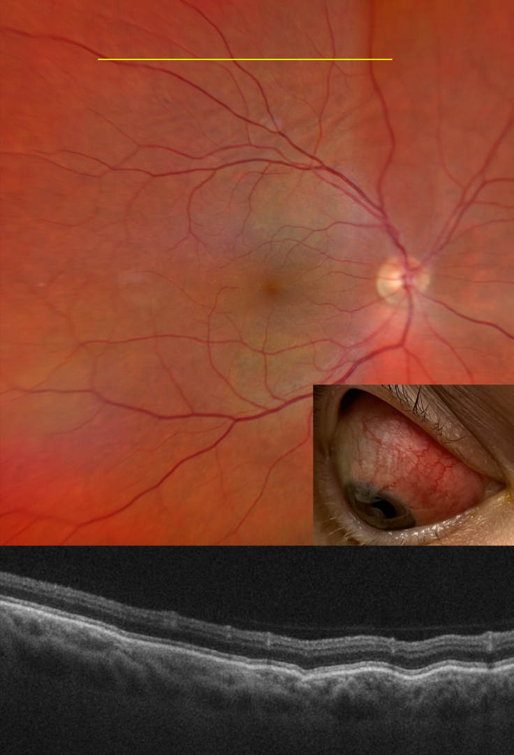
This 48YO female with a history of psoriatic arthritis presented on 10/25/22 with painful anterior scleritis. Vision was 20/20. The patient was started on 50mg of prednisone and slowly tapered off by rheumatology.
She returned on 7/19/23 with pain behind her right eye. Vision was 20/20. Color imaging shows subtle chorioretinal folds in the superior midperiphery.
OCT B-scan through this region confirmed the chorioretinal folds. B-scan ultrasonography revealed bilateral T signs.
Learning Points:
Posterior scleritis can present either as a diffuse thickening of the entire posterior sclera or as a nodular type with localized scleral thickening (see Agrawal et al, Retina 2016;36:392-401).
Nodular inflammation appears as a posterior pole subretinal yellowish mass lesion, often with overlying serous fluid and chorioretinal folds. B-scan ultrasonography may show fluid in the Tenon space (“T” sign).
Unlike anterior scleritis, the eyes are usually quiet and pain-free. Initial treatment is usually with oral non-steroidal anti-inflammatories or corticosteroids, although patients may also require immunosuppressive therapy. For a large case series of posterior scleritis, see McCluskey et al, Ophthalmology 1999;106:2380-2386.
Oral prednisone was restarted and slowly tapered. Further labs and chest X-ray were performed to rule out other possible causes of scleritis, and she was started on Humira.



