

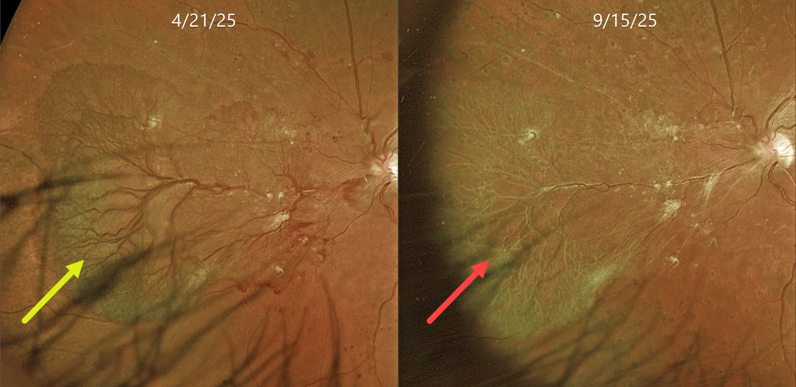
This 53YO female with a known history of type 2 diabetes was referred for asymptomatic proliferative disease (PDR) in her left eye. Vision was 20/50.
Optos color RG imaging shows a large sea fan-shaped area of succulent and flat neovascularization extending from the optic nerve into the nasal midperiphery. Panretinal photocoagulation (PRP) was applied in three sessions over several months.
Learning Points:
An attached vitreous provides the scaffold for neovascularization to grow onto and spread. Retinal neovascularization grows from the inner retinal surface and proliferates along the outer posterior hyaloid face (Vaz-Pereira et al, Retina 2017;37:1287-1296). Following PRP or anti-VEGF therapy for PDR, retinal neovascularization ‘involutes’, often morphing from a fine capillary network into larger vessels with fibrosis. In the process, the contracting neovascularization and vitreous can cause increasing traction. Rapid tissue contraction with traction retinal detachment is an uncommon but well-described phenomenon known as the ‘crunch’ syndrome (Survey of Ophthalmology 2021;66:926-932).
We were quite concerned about the possibility of a crunch in our patient and discussed it prior to starting treatment. Five months following her initial exam, the neovascularization completely fibrosed without traction. Vision was 20/40. We are continuing to follow her closely.



