

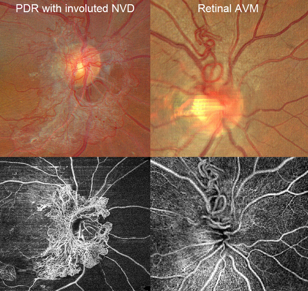
A patient with disc neovascularization (NVD) due to proliferative diabetic retinopathy (PDR) following full panretinal photocoagulation is compared with a patient with an asymptomatic retinal arteriovenous malformation (AVM).
Color photography of the PDR patient shows diffuse flat NVD with variable tight capillary networks of new vessels, admixed with larger vessels and fibrosis. OCT angiography (OCTA) confirms flow signals within these vessels, along with diffuse capillary loss superior, nasal, and inferior to the nerve. Color photography and OCTA of the retinal AVM show a tangle of arterioles and veins extending superiorly from the nerve. Fluorescein angiography, if performed, would show leakage from the NVD but not from the AVM.



