

This 28YO male with a history of type 1 diabetes was referred for asymptomatic retinal findings. He noticed some recent floaters in his right eye. Vision was 20/50 OU.
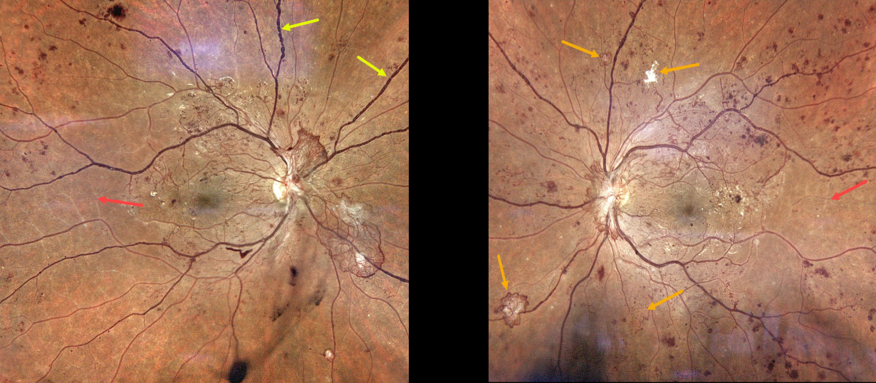
Color photography shows severe bilateral proliferative diabetic retinopathy. The right eye has a large area of active disc neovascularization, venous beading, and white vessels temporally indicating severe ischemia. There is some mild inferior vitreous blood.
The left eye has a smaller area of succulent NVD along with scattered smaller areas of variably active and fibrosed peripheral neovascularization. Clinical ischemic retina is also noted. Panretinal photocoagulation will be started bilaterally.



