

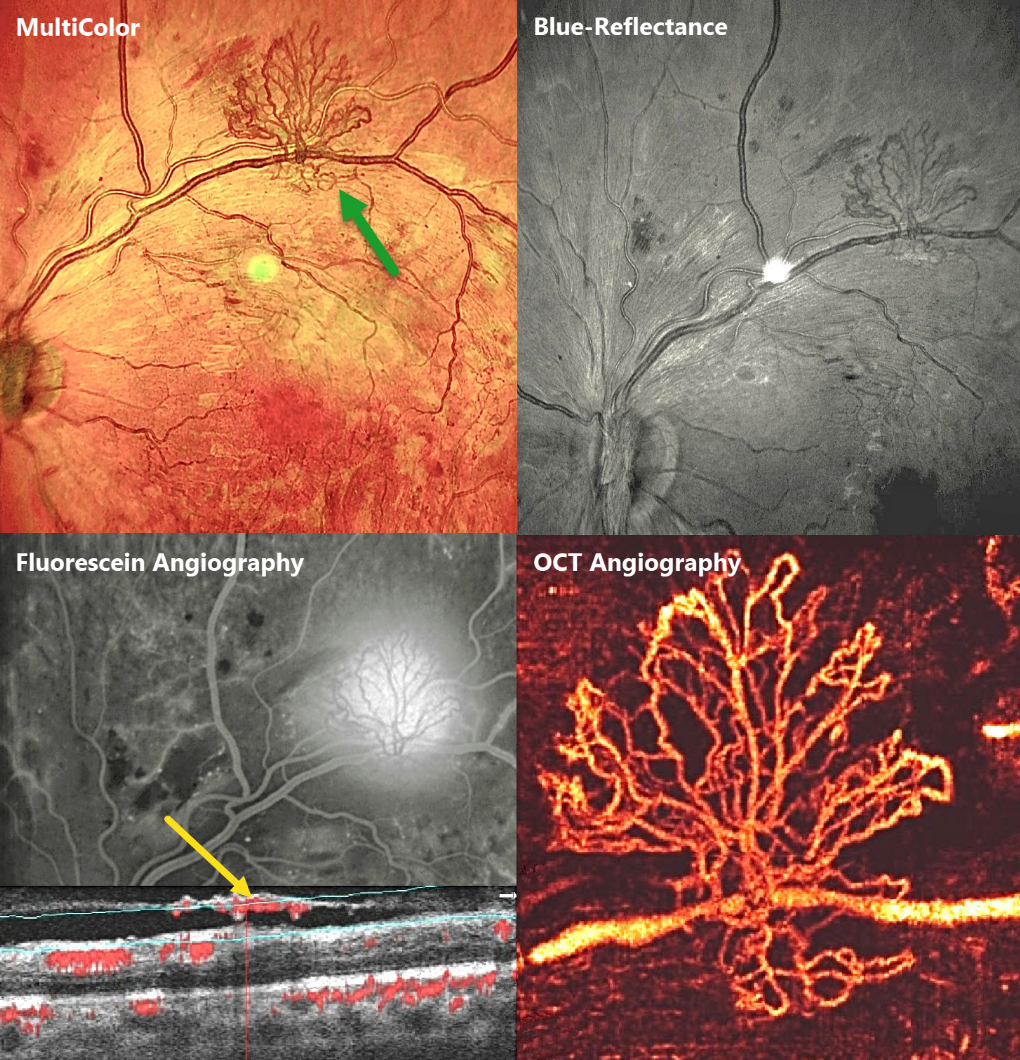
This 23YO male presented with bilateral proliferative diabetic retinopathy (PDR). Vision was 20/60 OD and 20/80 OS. Multimodal imaging of his left eye highlights an area of retinal neovascularization along the superotemporal arcade.
Which modality do you feel best images the neovascularization? Or are they all complementary?
MultiColor imaging shows the lacy active red vessels overlying the orange-red RPE and choroid. These vessels are better highlighted in the Blue-Reflectance image, since they appear black on a grey background. They profoundly leak on fluorescein angiography. En face OCT angiography (OCTA) beautifully shows the fine neovascular details. Finally, B-scan OCTA shows the neovascular vessels growing along the back surface of the partially detached posterior hyaloid.



