

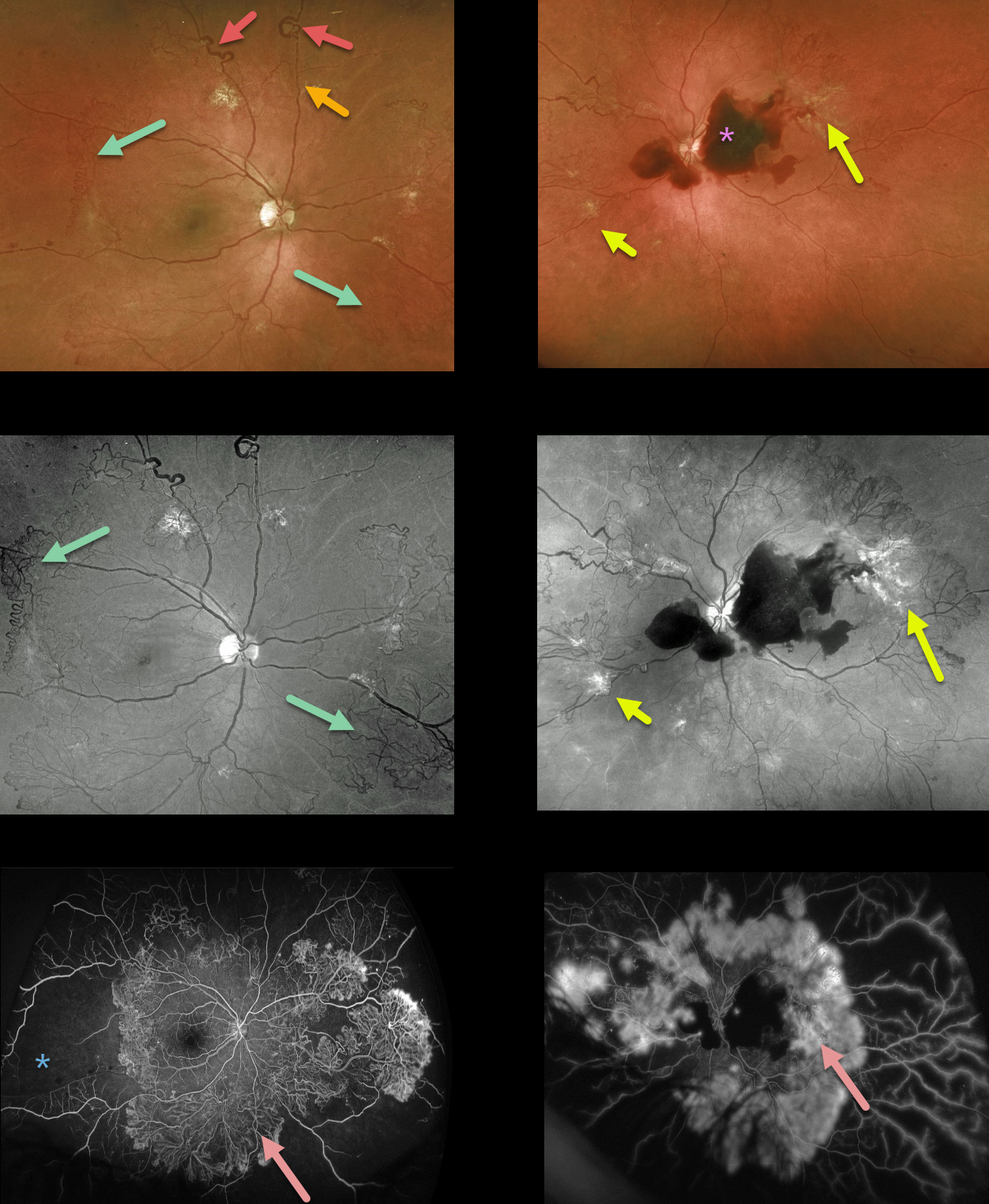
This 39YO presented with severe PDR in both eyes, highlighting nearly all of the key features of proliferative disease. Surprisingly, vision was 20/30 OD and 20/50 OS.
The right eye shows great examples of venous loops and venous beading and extensive scattered NVE, especially highlighted by the green channel Optos.
The left eye has a central subhyaloid hemorrhage along with venous beading and scattered NVE.
Fluorescein angiography shows dramatic and extensive neovascularization encircling both posterior poles with marked anterior ischemia.
Learning Points:
This case further emphasizes the importance of regular diabetic dilated examinations with peripheral retinal examination to unveil the true extent of the retinopathy.



