

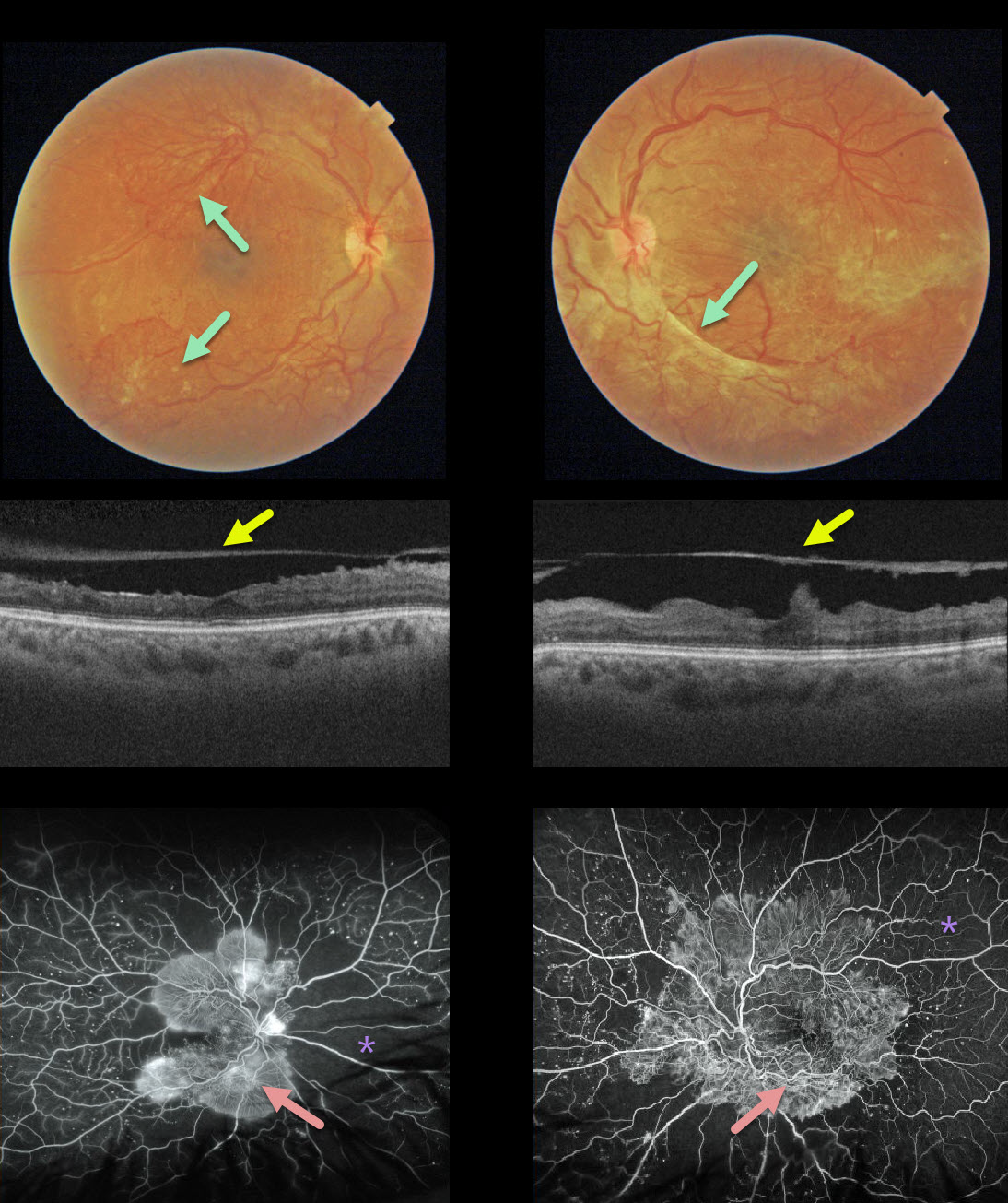
This 32YO female presented with 20/400 vision bilaterally, a hemoglobin A1C over 12%, and a blood glucose level over 300. Fibrosed neovascularization variably covers each posterior pole.
OCT shows that the posterior hyaloid has spontaneously detached from each central macula, which has a rippled contour. This appearance is also often seen following vitrectomy for tractional detachment with peeling of the hyaloid.
Fluorescein angiography shows extensive posterior pole neovascularization with peripheral ischemia.



