

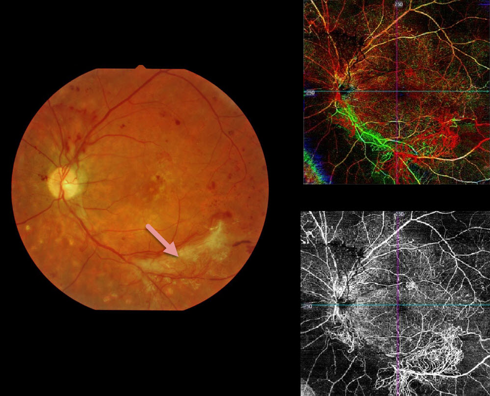
This patient has proliferative diabetic retinopathy. The fundus photo shows scattered red and fibrosed neovascularization.
The color OCTA image is a composite showing all layers of the retinal vasculature: red is the inner retina, green is the mid-retinal layer, and blue is the outer retina (photoreceptors, which are usually devoid of intrinsic vasculature). The bottom right image isolates the inner retina, showing regions of retinal ischemia with neovascularization.
Learning Points:
OCTA is a relatively new and non-invasive modality able to quickly identify diabetic retinal vascular abnormalities. Its role in screening for retinal ischemia and proliferative disease is still being defined.



