

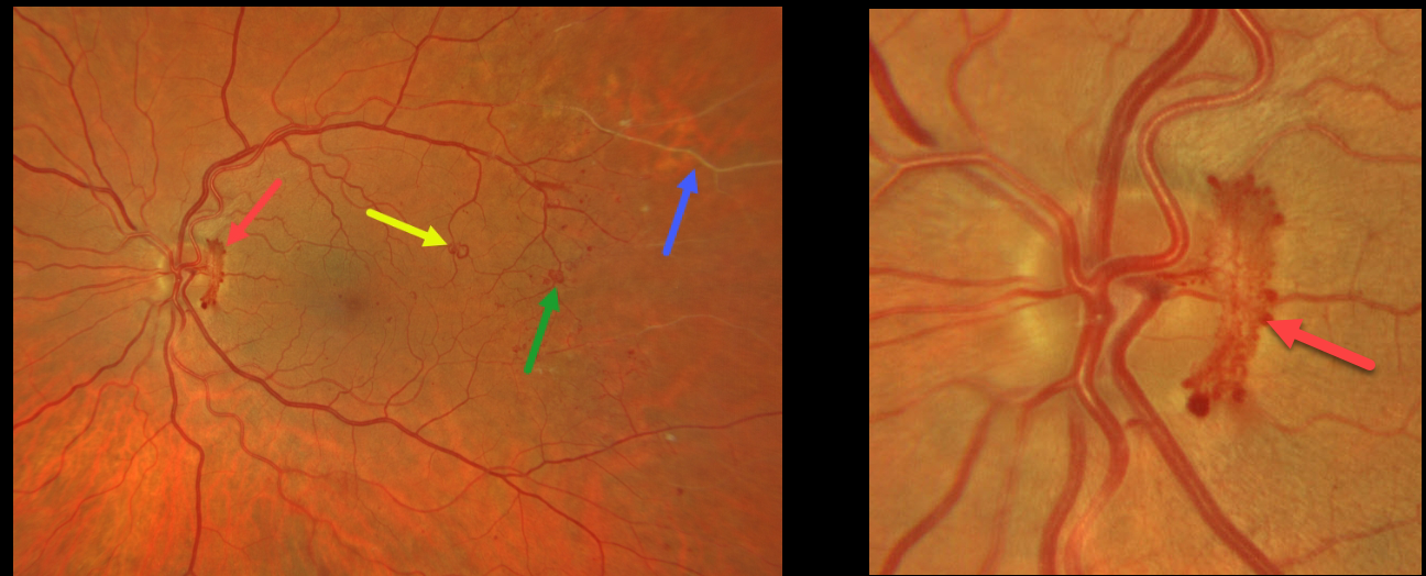
This patient presented with some beautifully imaged findings for proliferative diabetic retinopathy (PDR).
A wide-angle photograph shows active disc neovascularization (NVD), a venous loop, a few small patches of distal macular peripheral neovascularization (NVE), and distal retinal ischemia. A magnified image of the nerve shows the NVD.
Although venous loops are not generally included in the clinical classification of diabetic retinopathy, they are universally seen in severely ischemic eyes.
This patient is currently undergoing panretinal photocoagulation.



