

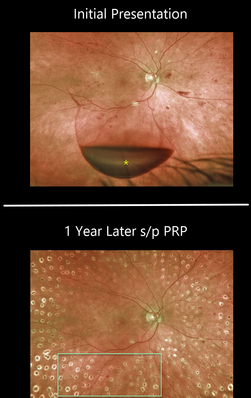
This 65YO female presented with 20/400 vision in her right eye. An acute inferior subhyaloid hemorrhage was noted along with non-central diabetic macular edema. Over the coming months, macular laser followed by panretinal photocoagulation (PRP) was performed.
One year later, vision was 20/50, with complete resolution of the preretinal blood and proliferative disease.
Learning Points:
Before PRP, patients with proliferative diabetic retinopathy (PDR) routinely went blind, and one of the treatments actually included pituitary ablation (for an amazing historical perspective on diabetic laser photocoagulation, see Goldberg and Jampol, Ophthalmology 1987;94;741-746).
PRP originally extended fairly posteriorly, and patients often needed fill-in treatment before vitrectomy and anti-VEGF treatments became available. Today, most laser surgeons will begin treatment well away from the nerve and major arcades to minimize the risks of central and peripheral vision loss.
Once considered a “one and done” treatment, more recent studies show that patients may still need additional laser, anti-VEGF injections, or vitrectomy (see Gonzalez et al, Ophthalmology 2021;128:1448-1457).
Some also advocate anti-VEGF injections as the primary treatment for PDR. However, most still prefer to add PRP at some point due to the risk of patients being lost to follow-up (see Gross et al, DRCR Protocol S, JAMA Ophthalmology 2018;136:1138-1148).



