

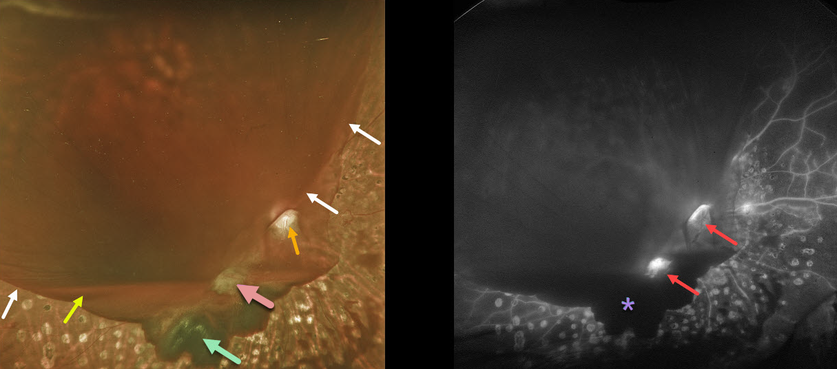
This diabetic patient developed a symptomatic hemorrhage in his right eye despite partial panretinal photocoagulation (PRP) treatment. The blood in our patient highlights the various layers of vitreous separation.
The reason why the far inferior and nasal retina and PRP scarring are visible is that the hyaloid is completely attached here. The hyaloid is shallowly elevated inferiorly, which gives a scalloped appearance to the dark red subhyaloid blood.
Elsewhere, the hyaloid is completely detached and highly elevated, allowing blood to pool in this space (white arrows mark the junction between the attached and detached hyaloid). A layer of blood pools inferiorly at the base of the hyaloid separation.
The optic disc and fibrosed neovascularization are well visualized because the vitreous remains tethered, preventing blood from accumulating.
Fluorescein angiography highlights areas of neovascularization and the blockage of the underlying retina by various layers of blood.
Learning Points:
At birth, the vitreous is diffusely adherent to the entire retina. But over the years, the vitreous begins to separate, first in the midperiphery, then in the peripheral macula, then the central macula, and finally the optic nerve.
For a great review of posterior vitreous detachment (PVD) evolution and complications, see Johnson AJO 2010;149:371-382.
Retinal neovascularization arises from the inner retina and extends onto the back surface of the vitreous; without this scaffold, it will not develop. A complete PVD is therefore protective against posterior segment retinal neovascularization.
Since the vitreous in the posterior pole is most adherent around the optic nerve and major arcades, this is where diabetic neovascularization most commonly develops. It is also why the vitreous often remains tethered to these structures as the proliferation progresses.
The posterior hyaloid continues to detach and contract, as does the neovascularization, which can cause vitreous hemorrhage and traction retinal detachment.



