

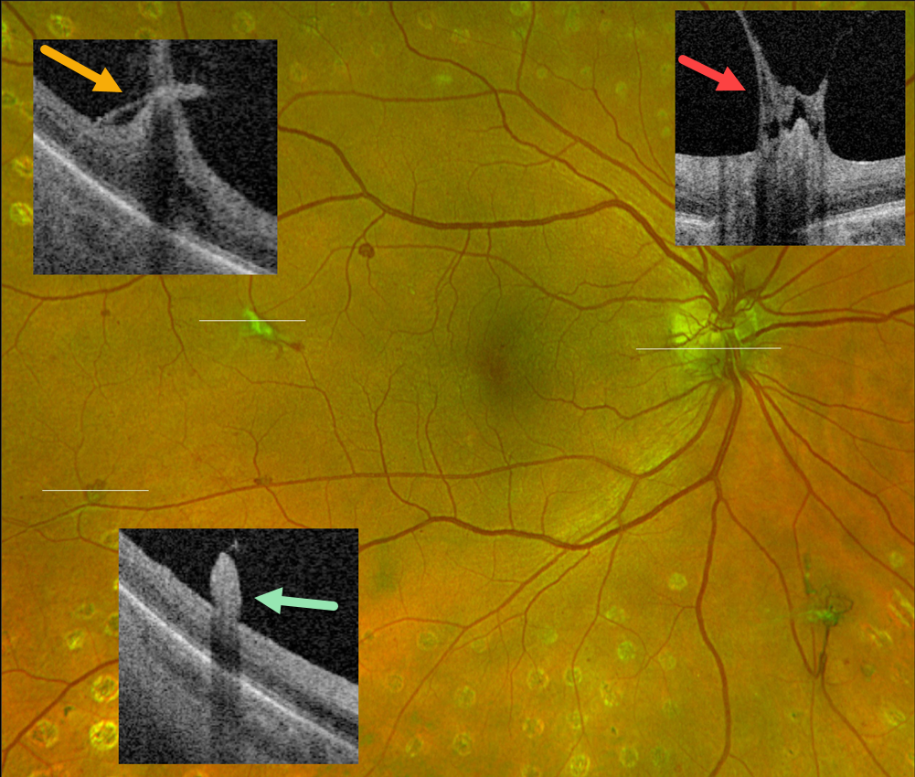
This 55YO female with type 2 diabetes has involuted proliferative diabetic retinopathy (PDR) following full panretinal photocoagulation.
Optos color RG imaging shows residual disc (NVD) and peripheral retinal neovascularization (NVE). Optos Silverstone swept-source OCT through disc and temporal macular neovascularization shows active vitreous traction on the residual vessels. There is no traction on small, reddish NVE in the distal inferotemporal macula. Continued observation was recommended.
Learning Points:
An attached vitreous provides the scaffold for neovascularization to grow onto and spread. Retinal neovascularization grows from the inner retinal surface and proliferates along the outer posterior hyaloid face (Vaz-Pereira et al, Retina 2017;37:1287-1296). This tethering to a partially detached and contracting hyaloid is what causes tractional retinal detachment and vitreous hemorrhage. Thus, although an eye with a complete posterior vitreous detachment can develop small buds of neovascularization along the retinal surface (as seen in our patient’s inferotemporal NVE), the detached hyaloid is protective against the growth of large sheets of fibrovascular tissue.



