

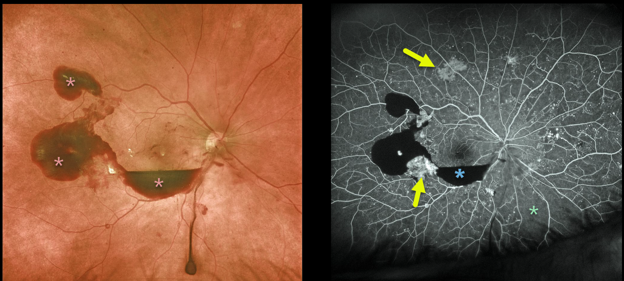
The 58 yo male with proliferative diabetic retinopathy presented with preretinal blood. Optos fluorescein angiography shows blockage from this blood with scattered areas of midperipheral neovascularization and peripheral ischemia, especially inferonasally.
Learning Points:
Angiographically, hypofluorescence is classified as either a blockage or a filling defect. Blood, which masks the underlying retinal and choroidal fluorescence, causes blockage. There is diffuse capillary loss and ischemia, appearing as glassy-dark areas of hypofluorescence throughout, representing filling defects. Filling defects are virtually never as dark as areas of blockage.
Angiographic hyperfluorescence is classified into window defects, pooling, staining, leakage, and neovascularization. The new blood vessels adjacent to the subhyaloid blood and in the mid-periphery lack endothelial tight junctions, which is why neovascularization shows profound angiographic leakage.



