

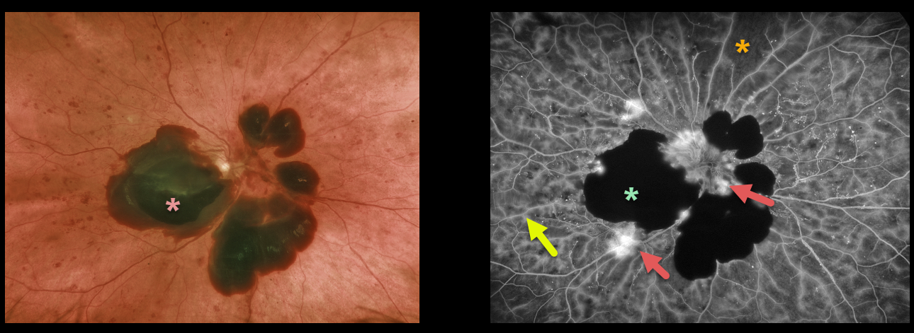
This 53YO female with type 2 diabetes presented with acute vision loss from previously undiagnosed proliferative diabetic retinopathy.
Optos color imaging shows patches of mostly subhyaloid blood in the macula and peripapillary retina.
Fluorescein angiography beautifully exhibits various angiographic characteristics for hypo- and hyperfluorescence. We can see both blocking and filling defects, in addition to diffuse vascular staining and leakage of the disc and adjacent to the subhyaloid blood.
Learning Points:
Angiographically, hypofluorescence is classified as either a blockage or a filling defect. Blood, which masks the underlying retinal and choroidal fluorescence, causes blockage. There is diffuse capillary loss and ischemia, appearing as glassy-dark areas of hypofluorescence throughout, representing filling defects. Filling defects are virtually never as dark as areas of blockage.
Angiographic hyperfluorescence is classified into window defects, pooling, staining, leakage, and neovascularization, and our case exhibits several of these features.
The endothelial cells of the larger vessels in areas of capillary dropout become ischemic, leading to breakdown of the inner retinal barrier. The larger vessels, therefore, often show late staining in these regions as fluorescein dye leaks into and stains the vessel wall. Vascular staining is evident throughout the entire imaged fundus.
The new blood vessels growing on the disc and adjacent to the subhyaloid blood lack endothelial tight junctions, which is why neovascularization shows profound angiographic leakage.



