

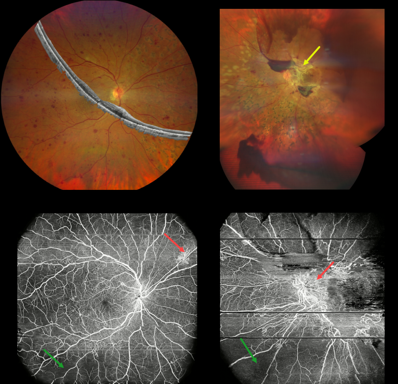
This 43YO female has a 16-year history of type 1 diabetes, and presented with 3 years of progressive vision loss in her left eye. Vision was 20/20 OD and 20/400 OS.
Composite widefield color imaging and OCT of her right eye show moderate retinal hemorrhages, nasal panretinal photocoagulation scarring, and no edema. Widefield OCT angiography shows peripheral ischemia with superonasal midperipheral neovascularization.
Multimodal imaging of her left eye shows fibrotic neovascularization around the nerve and macula. OCT angiography shows the neovascularization and peripheral retinal ischemia.
Learning Points:
Ultra-widefield imaging with fluorescein angiography is becoming increasingly important in screening for proliferative disease, especially since about 70% of angiographic nonperfusion lies outside the posterior pole (Silva et al, DRCR Protocol AA, Retina 2022;42:1302-1310). Widefield OCT angiography may also prove to be an equally effective yet less invasive screening tool (Al-Khersan et al., Am J Ophthalmol. 2021;224:292-300).



