

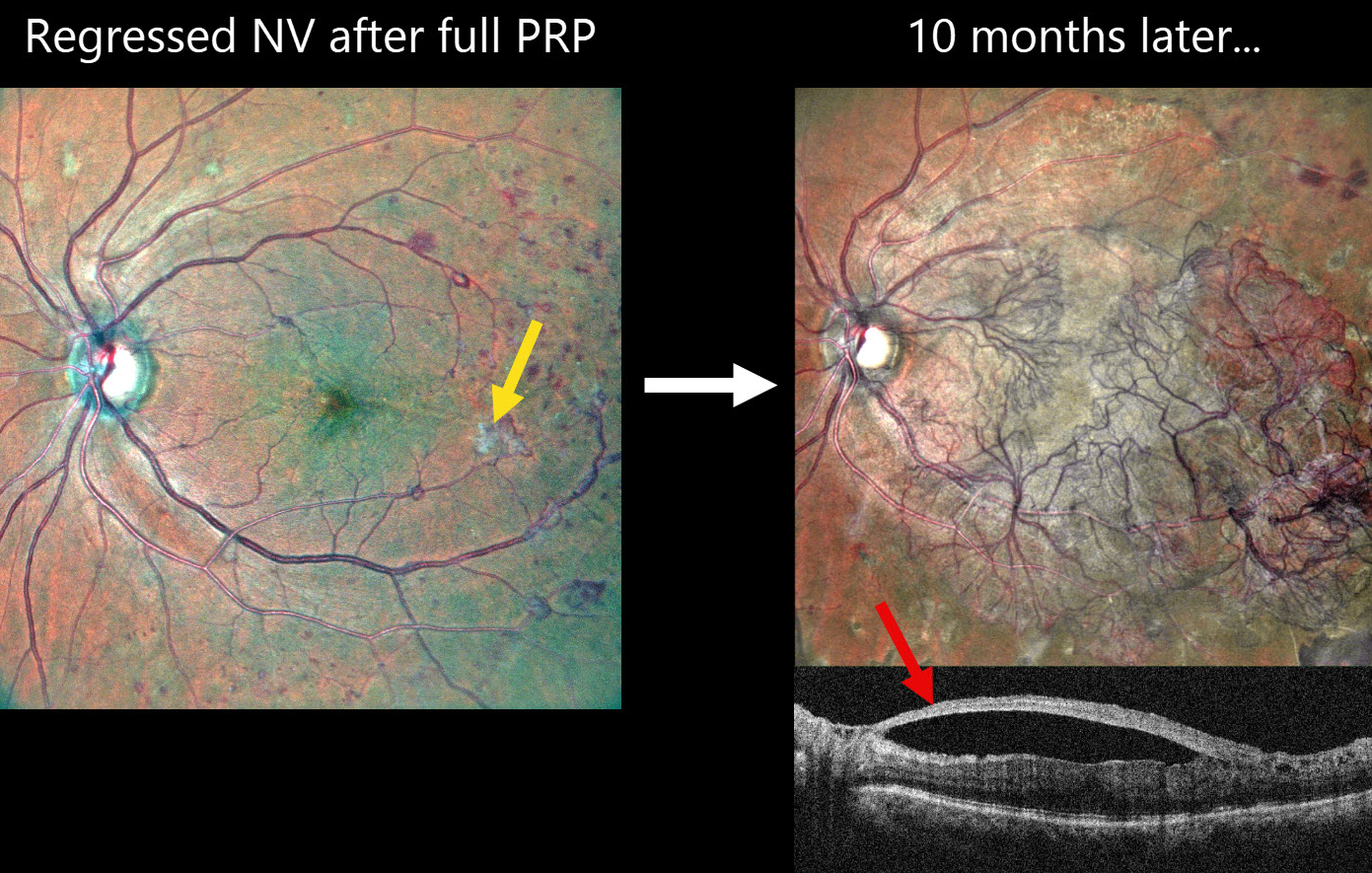
This 60YO female with a history of type 2 diabetes underwent full panretinal photocoagulation (PRP) in her left eye. Following laser, MultiColor SLO imaging shows small residual areas of variably fibrosed retinal neovascularization. Vision was 20/30.
Ten months later, she presented complaining of 2 months of decreased vision in this eye. Vision was 20/60. MultiColor SLO imaging shows retinal neovascularization covering the posterior pole. On OCT, the fovea is remarkably attached with the hyperreflective neovascularization elevated along the partially detached posterior hyaloid.
Learning Points:
Although PRP is often considered a ‘one and done’ procedure, this is not always the case (Gonzalez et al, Ophthalmology 2021;128:1448-1457). Our patient is a perfect example of why these patients require lifelong monitoring even if their disease initially appears quiescent.
Given the aggressive neovascular proliferation, intravitreal anti-VEGF was given, followed by vitrectomy 5 days later. Although PRP is often considered a ‘one and done’ procedure, this is not always the case (Gonzalez et al, Ophthalmology 2021;128:1448-1457). Our patient is a perfect example of why these patients require lifelong monitoring even if their disease initially appears quiescent.
Given the aggressive neovascular proliferation, intravitreal anti-VEGF was given, followed by vitrectomy 5 days later.



