

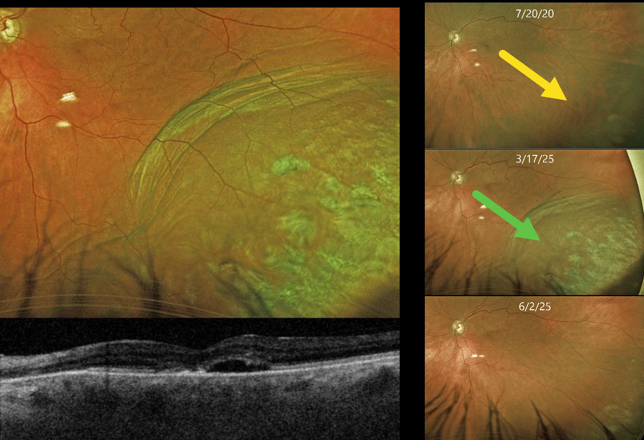
This 85YO female presented with an elevated pigmented choroidal mass in her inferotemporal periphery, which extended into her distal macula (Optos color RG). This mass was absent 5 years earlier. Circumferential choroidal folds extend along the posterior edge of the lesion. Triton swept-source OCT shows a blister of subretinal fluid and a thickened choroid.
She has a history of uncontrolled glaucoma and underwent a XEN Gel Stent procedure 8 days earlier. Intraocular pressure (IOP) was 8 mmHg.
Learning Points:
A choroidal detachment is defined by the abnormal presence of fluid or blood in the suprachoroidal space. Serous choroidal detachments, also known as choroidal effusions, are a frequent complication of glaucoma surgery. Several causative mechanisms are believed to be at play: hypotony allows fluid to accumulate in the suprachoroidal space, and inflammation increases the choroidal permeability. The detached ciliary body may also produce less aqueous, thus creating a self-perpetuating cycle. Often, observation is sufficient, but topical steroids and cycloplegics can be helpful in promoting resolution.
Other than the obvious appearance of this lesion immediately following glaucoma surgery, clues to this not being a uveal MM include the choroidal folds along the posterior extent of the choroidal detachment and an OCT consistent with hypotony maculopathy. About 10 weeks later, the IOP was 9 mmHg, and the choroidal effusion had completely resolved.



