

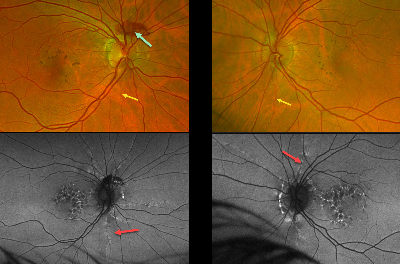
This 53YO female was referred for asymptomatic macular findings. Her mother has a history of pseudoxanthoma elasticum (PXE). Vision was 20/25 OD and 20/40 OS.
Optos color RG imaging shows coarse subretinal pigment clumps bilaterally. Subretinal blood is noted along the superior aspect of the right nerve from a peripapillary choroidal neovascularization. A few barely visible angioid streaks radiate from each nerve.
Fundus autofluorescence (FAF) shows variable hyper- and hypo-FAF flecks. The angioid streaks appear as hyper-FAF lesions radiating outwards from each nerve.
Learning Points:
Retinal findings in PXE most commonly include angioid streaks and a subretinal peaux d’orange appearance, most pronounced in each temporal macula. However, pattern dystrophy-like changes, as in our patient, can sometimes be the predominant phenotype (Murro et al, Graefe’s 2020;258:1881-1892). As with typical pattern dystrophies, the findings are dynamic and best captured with multimodal imaging.
PXE is caused by mutations in the ABCA6 gene, and our patient tested positive for homozygous pathogenic ABCA6 mutations. PXE is virtually always transmitted as an autosomal recessive disorder, although autosomal dominant transmission may rarely occur (Plomp et al, Am J Med Genet A 2004;126A:403-412).



