

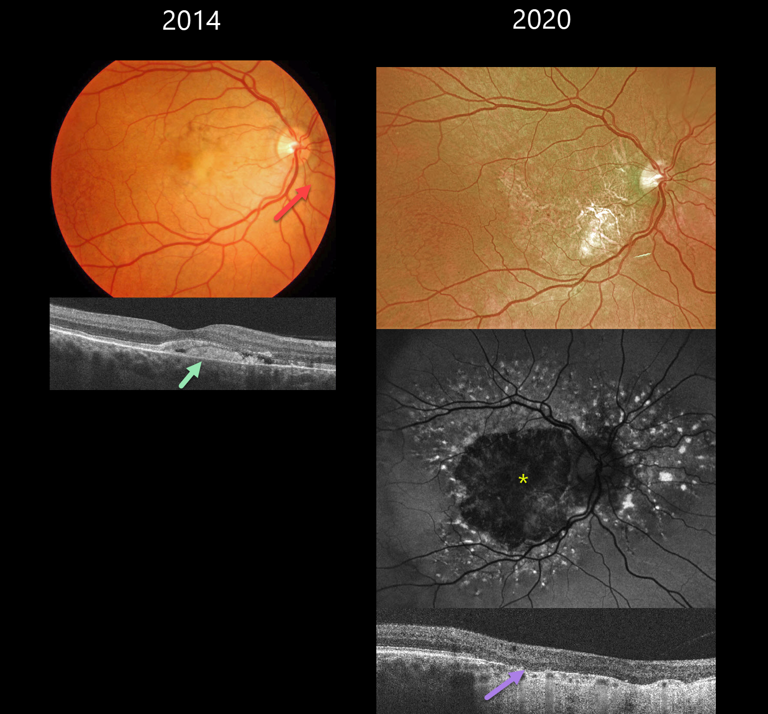
This 39YO male with a known history of pseudoxanthoma elasticum presented on 2/19/14. Findings were bilateral at this, and subsequent visits, so only his right eye images will be shown and discussed.
Color photography shows nasal yellowish subfoveal pigmentary changes with a peaux d’orange appearance temporally. Some peripapillary angioid streaks are noted. OCT shows hyperreflective vitelliform material and fluid below the retina, with some of this material also infiltrating the outer retina.
He has been followed without treatment, and when examined on 7/17/20, vision was 20/200 OD and 20/80 OS. Optos color RG imaging shows a variably atrophic macula. The atrophy on fundus autofluorescence is hypo-FAF. There is a fairly dramatic pattern of hyper-FAF spots radiating out from this atrophy. OCT shows outer retinal atrophy and no fluid. When last examined on 7/6/23, vision was 20/200 OU.
Learning Points:
Retinal findings in PXE most commonly include angioid streaks and a subretinal orange skin (peaux d’orange) appearance, most pronounced in each temporal macula. However, pattern dystrophy-like changes, as in our patient, can sometimes be the predominant phenotype (Murro et al, Graefe’s 2020;258:1881-1892). As with typical pattern dystrophies, the findings are dynamic and best captured with multimodal imaging.



