

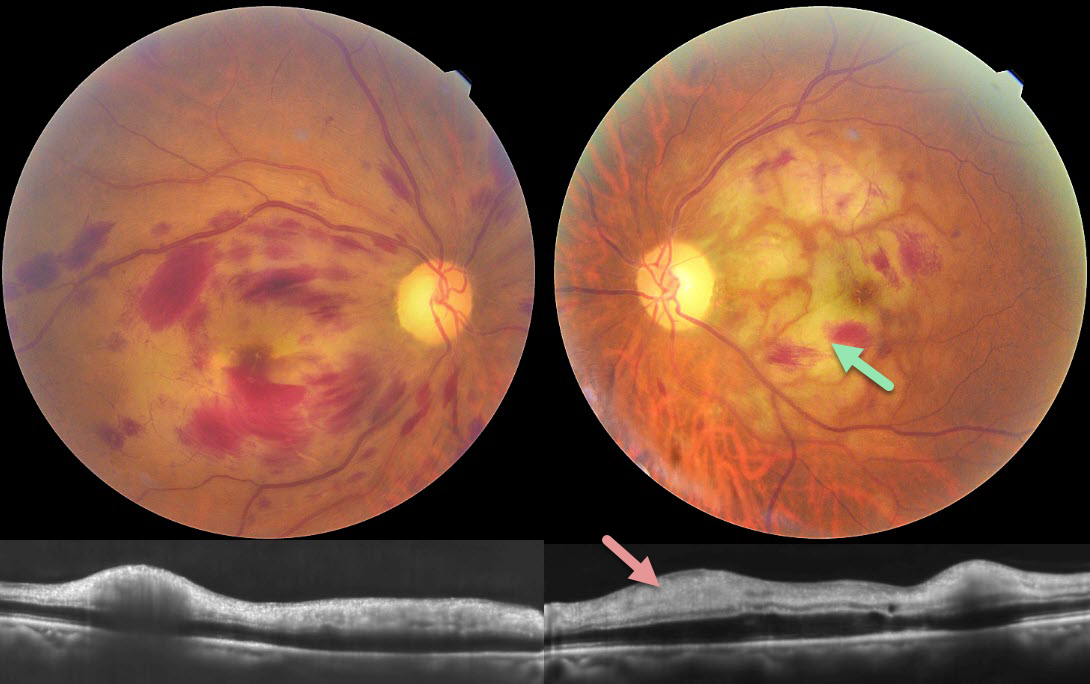
This 85YO male lost vision in each eye following a fall around 1 week back with multiple rib fractures. Vision was hand motion OU.
Color photography shows bilateral macular retinal hemorrhages with patches of white ischemic retina, most prominent in the left macula. OCT scanning shows a thickened hyperreflective inner retina bilaterally.
Learning Points:
Purtscher retinopathy was first described by Otmar Purtscher in 1910. Although originally described in a man who fell from a tree with cranial trauma, these white patches of retinal ischemia and hemorrhages are found in numerous other etiologies, including pancreatitis, chest trauma, and collagen vascular diseases. The findings are called Purtscher retinopathy when due to trauma (as in our patient), and Purtscher-like retinopathy when seen from other causes.
The underlying cause is unknown, but the findings are thought to result from leukoembolization due to complement activation. The areas of ischemia include more superficial nerve fiber layer infarcts (cotton wool spots) and deeper ischemia involving the middle retinal layers.



