

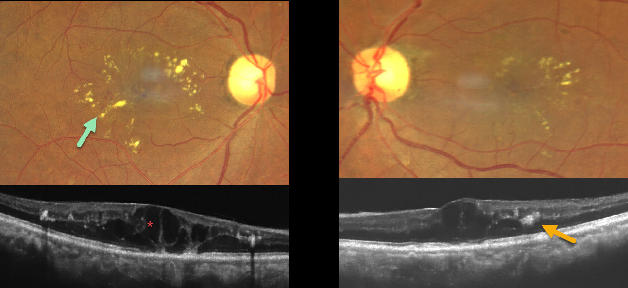
This 53YO female presented with gradual bilateral vision loss. Three years earlier, she was treated elsewhere for metastatic breast carcinoma, which included a mastectomy followed by radiotherapy to the supraclavicular region and brain. She denied having diabetes. Vision was 20/60 OD and 20/30 OS.
Fundus photography shows bilateral macular telangiectasia with radiating macular lipid flecks. OCT scanning shows diffuse cystic retinal thickening with hyperreflective lipid centered around the outer plexiform layer. Anti-VEGF therapy was recommended.
Learning Points:
Radiation retinopathy usually occurs 3 months to 3 years after external-beam radiation or plaque radiotherapy. The ocular findings and pathogenesis closely resemble those seen with diabetic retinopathy. Radiation optic neuropathy can also develop (Carey et al, Br J Ophthalmol 2023;107:743-749).



