

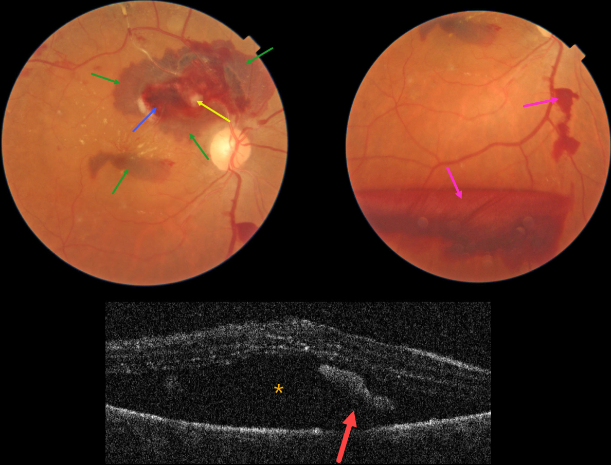
This 76YO female presented with blurred vision in her right eye for one month. Vision was 20/60 in her right eye and 20/30 in her left eye.
Color photography shows a ruptured retinal arterial macroaneurysm (RAM) causing multiple layers of blood. The blood is subretinal, intraretinal, and preretinal. Some scattered lipid flecks are noted centrally and temporally, and the arterial distal to the RAM is sclerosed.
OCT scanning shows marked subretinal fluid and some hyperreflective subretinal blood.
Learning Points:
A RAM is thought to occur in a weakened arterial wall from arteriosclerosis, and appears as a yellow-white dilated lesion along a retinal artery within the first 3 bifurcations from the optic nerve.
There are 2 distinct presentations, hemorrhagic or exudative. The hemorrhagic type often presents, as in this case, with pathognomonic multiple layers of blood. Acute macular exudation can be treated with observation, anti-VEGF injections, or thermal laser.
Due to her symptomatic subretinal fluid, a single intravitreal Avastin injection was given. Four months later, vision improved to 20/30, the macular fluid resolved, and most of the blood layers were gone.



