

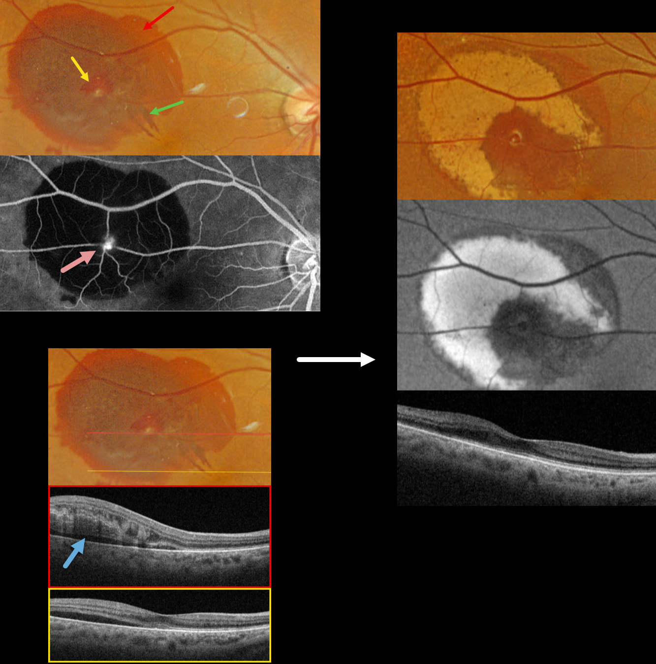
This 83YO female presented with recent blurred vision in her right eye. Vision was 20/40.
Optos color RGB imaging shows subretinal blood in the superotemporal macula. A small amount of inner vs preretinal blood surrounds a small yellow-white retinal arterial macroaneurysm (RAM). In addition, some radiating outer plexiform blood radiates towards the macular center. Fluorescein angiography shows a hyperreflective, leaking RAM with blockage by surrounding subretinal blood. Swept source OCT shows variable outer retinal edema, hyperreflective blood, and subretinal fluid.
Observation was recommended. Six weeks later, the blood is absorbing and turning yellow superiorly and temporally. This devitalized blood is markedly hyper-autofluorescent. The foveal fluid resolved, and vision improved to 20/30.
Learning Points:
A RAM is thought to occur in a weakened arterial wall from arteriosclerosis and appears as a yellow-white dilated lesion along a retinal arterial within the first 3 bifurcations from the optic nerve. There are 2 distinct presentations, hemorrhagic or exudative. The hemorrhagic type often presents, as in this case, with pathognomonic multiple layers of blood. Acute macular exudation can be treated with observation, anti-VEGF injections, or thermal laser



