

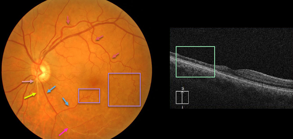
At first or quick glance, this looks like an old inferotemporal branch retinal vein occlusion (BRVO).
On closer inspection, there is a white-sheathed vessel arising from the nerve inferiorly. Numerous collateral vessels are noted inferior and temporal to the fovea, and there is a sheathed vessel in the inferior arcade.
OCT shows marked inner retinal atrophy with preservation of the photoreceptor layers, consistent with a prior branch retinal artery occlusion. So the collateral vessels (virtually always a sign of a prior venous occlusion) indicate an old BRVO, and the atrophic and sheathed vessel indicates an old BRAO, both in the same quadrant. Right?
Tracing the large branch of the superotemporal arcade inferotemporally, we see that the collaterals appear to be arterial. Inferior to the nerve, we see an arteriole that crosses the sheathed vessel, indicating that the sheathed vessel is not an artery but a vein. Moreover, the large vessel off the nerve, inferotemporal in location, appears to be a vein that eventually connects to the collateral vessels we previously identified as arterial.
Our best guess is that this may be a low-grade arteriovenous malformation, possibly with a prior secondary BRAO/BRVO. Unfortunately, we don’t have a fluorescein angiogram, which may help better define the vascularization.
Learning Points:
Remember that arteries don’t cross arteries and veins don’t cross veins. Tracing vessels can be a helpful key to identifying retinal vascular disorders.



