

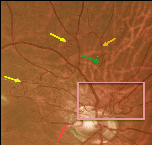
This 61YO male presented with these asymptomatic blood vessels extending superiorly from the right nerve. Vision was 20/25 OU, and the left eye was normal.
The abnormal vessels emanate from a cilioretinal artery, extend superiorly into and then above the macula, then arch nasally and descend. Note that it crosses a vein, confirming that it is still arterial (remember that arteries cross veins, veins cross arteries, but arteries never cross arteries and veins never cross veins). It then descends towards the nerve in a tangle of vessels likely consisting of both arteries and veins.
We felt that these vessels represented a low-grade arteriovenous malformation (AVM).
Learning Points:
Since about 25% of patients with AVMs can have intracranial venous anomalies, a brain MRI is usually recommended.
These vessels somewhat resemble collateral vessels noted after retinal vein occlusions. Collaterals are dilated capillaries that result from increased hydrostatic pressure from an occlusion. They are almost always venous, and when seen, are a telltale sign for a prior occlusion. Unlike retinal neovascularization, they do not leak angiographically.



