

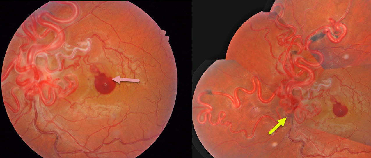
This healthy 34YO man noticed sudden vision loss in his left eye following heavy lifting 2 days earlier. Vision was 20/20 in his normal OD and 20/200 OS.
Color photography shows a diffuse network of markedly dilated, tortuous vessels involving the optic disc and posterior pole, extending into the peripheral retina, with an associated small premacular hemorrhage. The vessels are sheathed superonasal to this blood. Unfortunately, our patient was lost to follow-up immediately.
Learning Points:
A retinal arteriovenous malformation (AVM) is a congenital connection between an artery and a vein without an intervening capillary network. These lesions appear along a spectrum ranging from small, isolated lesions to the large ‘bag of worms’ appearance. These more extensive AVMs are usually part of the Wyburn-Mason Syndrome, which includes ocular and midbrain vascular lesions. Although retinal AVMs are usually stable lesions, bleeding or secondary venous occlusions may occur. Our patient’s vascular sheathing may indicate prior occlusion.



