

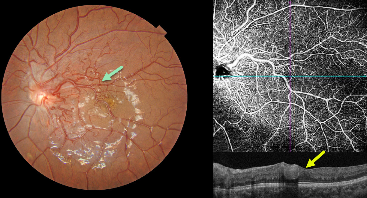
This 10YO girl presented with asymptomatic retinal findings in her left eye. Vision was 20/25 in her normal OD and 20/30 OS.
Color photography shows a retinal arteriovenous malformation (AVM) composed of dilated, tortuous vessels, mostly in the macular area. The foveal pit is crossed by a large tortuous vessel. OCT angiography highlights the profoundly abnormal macular vasculature, and an OCT B-scan shows the markedly dilated inner foveal vessel. MRI scanning of the brain and orbits was normal.
Learning Points:
A retinal AVM is a congenital connection between an artery and a vein without an intervening capillary network. An AVM appears along a spectrum ranging from small, isolated lesions to the large ‘bag of worms’ appearance. These more extensive lesions are usually part of the Wyburn-Mason Syndrome, which includes ocular and midbrain vascular lesions. Our patient has an isolated retinal AVM, as there are no intracranial lesions.



