

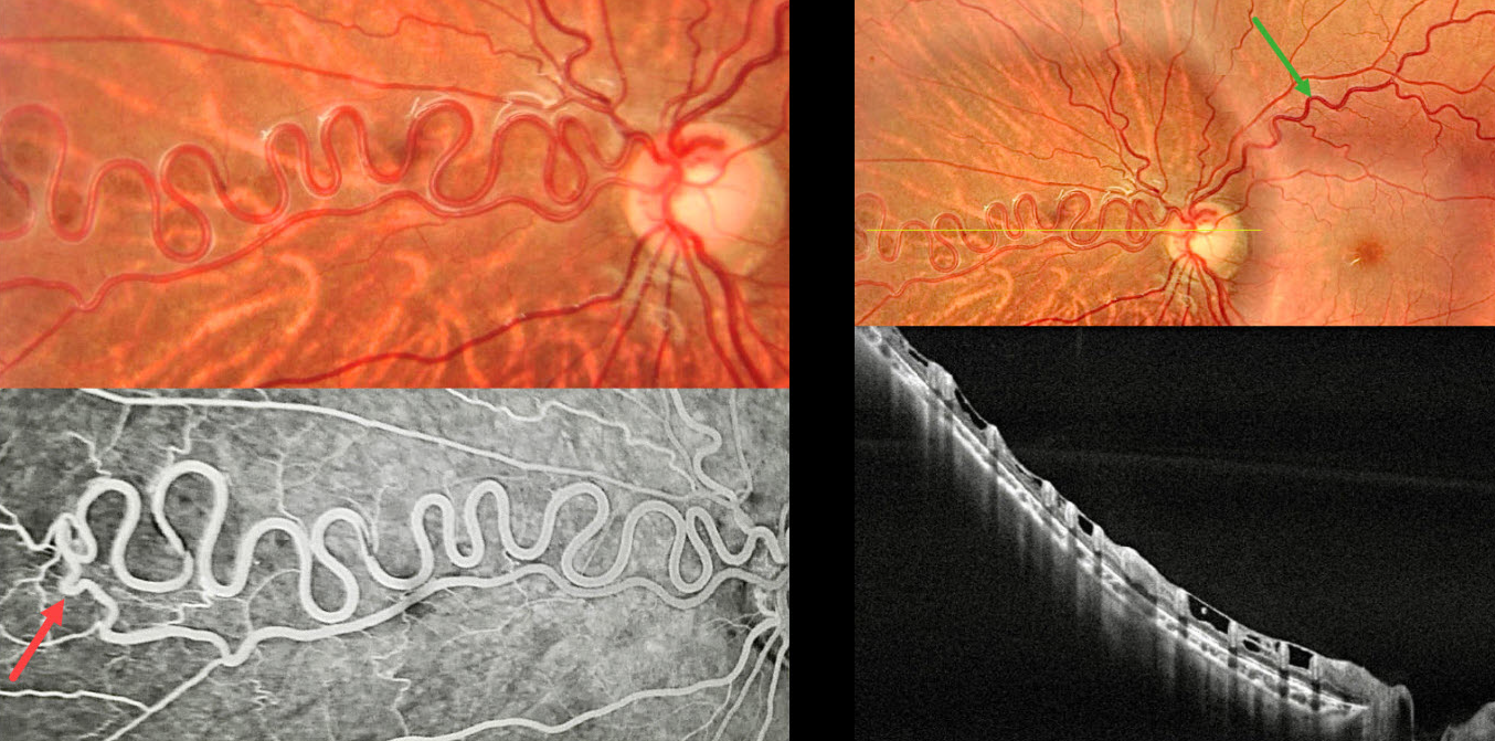
This healthy 12YO boy was referred for asymptomatic retinal findings. Vision was 20/20 in his normal OD and 20/20 OS.
Color photography shows a dilated multi-corkscrewed arteriole extending into the nasal midperiphery. The major superotemporal vein is also somewhat dilated and tortuous. On OCT, these anomalous vessels are hyperreflective and extend through the full thickness of the retina. ILM draping spans between each vessel. Angiographically, the artery directly connects to a paired vein. There is no angiographic leakage.
Learning Points:
A retinal arteriovenous malformation (AVM) is a congenital connection between an artery and a vein without an intervening capillary network. These lesions appear along a spectrum ranging from small, isolated lesions to the large ‘bag of worms’ appearance. These more extensive lesions are usually part of the Wyburn-Mason Syndrome, which includes ocular and midbrain vascular lesions. Although retinal AVMs are usually stable lesions, secondary venous occlusions may occur. Our patient has an isolated retinal AVM due to the lack of intracranial lesions on neurologic imaging.



