

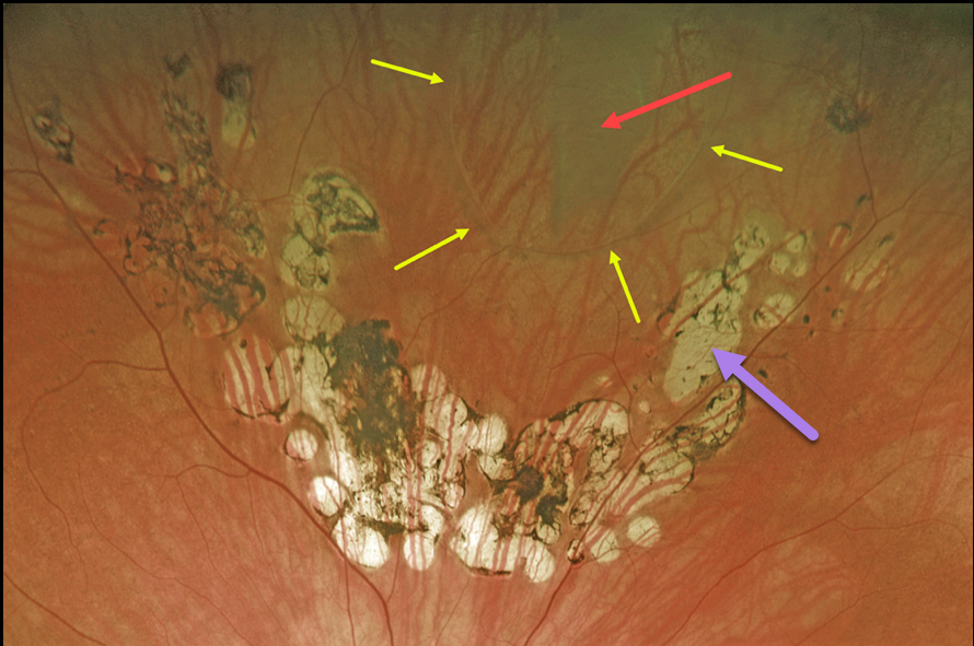
This 67YO female presented with a symptomatic vitreous floater that started about 6 months earlier when she developed a symptomatic retinal tear.
The break was treated elsewhere with laser photocoagulation, and on subsequent visits, she was told the treatment was successful and looked perfect.
However, Optos imaging in our office shows otherwise. A large tear is visualized superiorly. Laser scars are seen posterior to the tear, but barely extend up its lateral margins and completely miss the anterior edge. Even where present, the scarring doesn’t extend to the edge of the break.
Elective vitrectomy surgery will be performed in the near future for her symptomatic vitreous floaters.
We will complete the prophylactic retinopexy at the time of surgery.
Learning Points:
Symptomatic retinal tears in the presence of an acute posterior vitreous detachment have a high risk of causing a rhegmatogenous retinal detachment. Prophylactic retinopexy is therefore indicated.
Treatment must completely surround the tear and, ideally, extend to the ora serrata. Both thermal laser photocoagulation and cryotherapy are equally effective in creating a vitreoretinal adhesion, which takes at least several weeks to form.



