

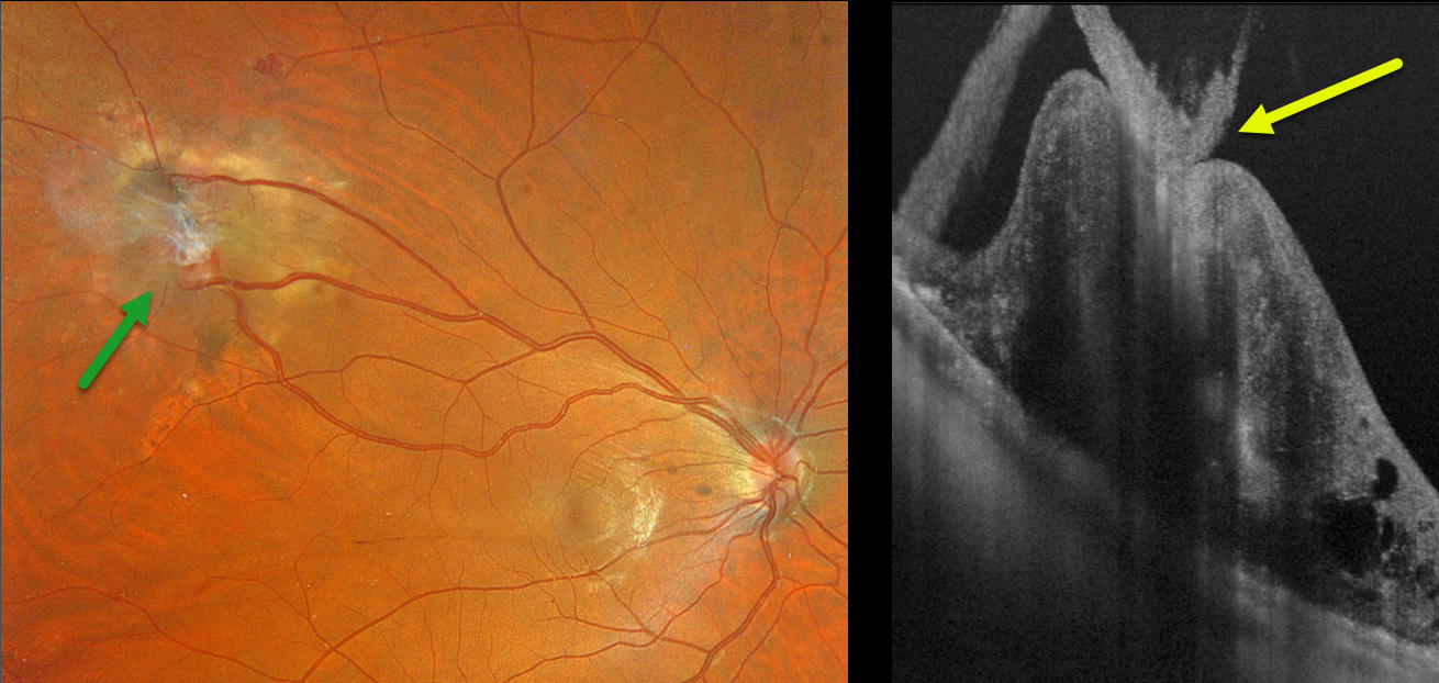
This 28YO male with a known history of Von Hippel-Lindau (VHL) recently moved to our area. He received prior anti-VEGF injections and thermal laser for a retinal capillary hemangioma in his right eye.
Optos color RGB imaging shows a regressed endophytic capillary hemangioma in the superotemporal midperiphery. The tumor is supplied and drained by dilated, irregular vessels. On Silverstone swept-source OCT, the angioma appears as a thickened, hyperreflective lesion with overlying vitreous traction.
Learning Points:
Retinal capillary hemangiomas are orange-red lesions associated with a prominent paired feeding and draining vessel and can cause severe vision loss or blindness from exudative retinal detachment. These benign vascular tumors need to be aggressively treated. Common treatment options include thermal laser, cryotherapy, and anti-VEGF injections.
Retinal capillary hemangioma is a predominant feature of VHL, an autosomal dominant condition caused by mutations in the VHL tumor suppressor gene. Patients develop benign and malignant tumors, including central nervous system hemangioblastoma, pheochromocytoma, and renal cell carcinoma.
We plan on following our patient several times per year to monitor for new lesions or recurrent exudation.



