

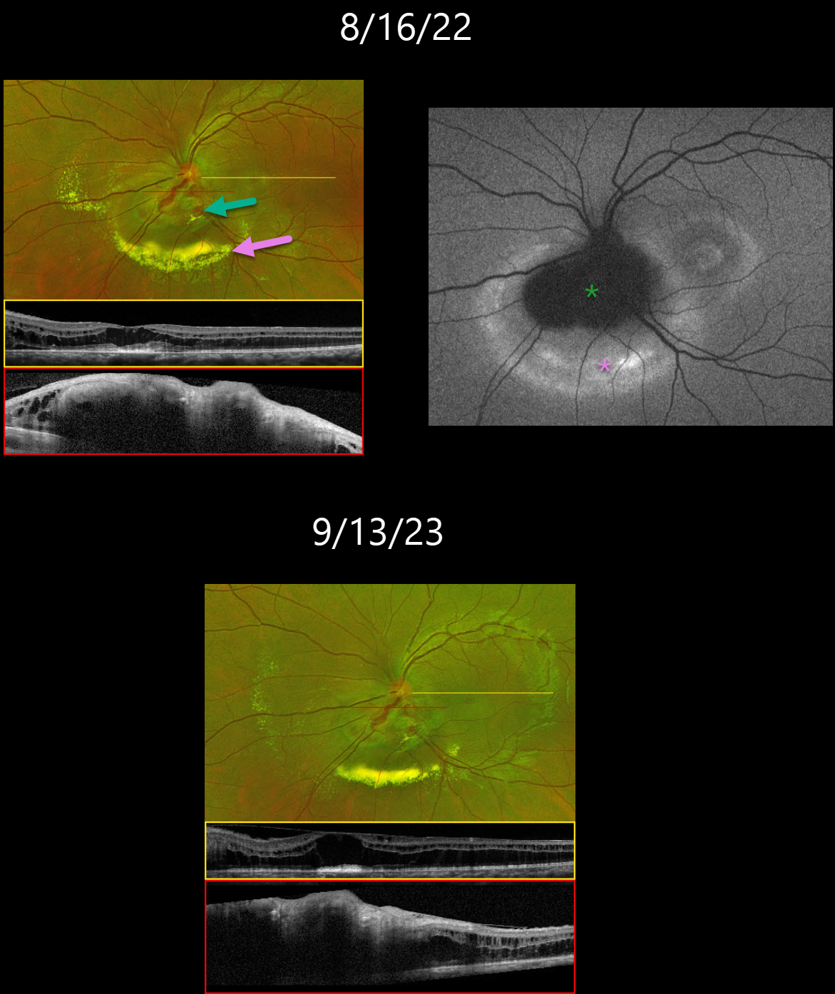
This 23YO female was noted by her optometrist in 2019 to have vision loss from a retinal lesion. At presentation, vision was 20/400 in her left eye and 20/20 in the right eye.
She was referred to an outside retina specialist, who gave a single anti-VEGF injection and then referred her to Columbia University. Our initial examination on 8/16/22 revealed an inferior peripapillary retinal capillary hemangioma with surrounding exudation. OCT of the macula revealed mostly outer retinal fluid. The angioma was diffusely thickened and hyperreflective.
On fundus autofluorescence (FAF), the angioma was hypo-FAF with a surrounding rim of hyper-FAF. Genetic testing for the VHL gene was negative, and thus further systemic work-up of the brain and kidneys was deferred.
She underwent subsequent monthly anti-VEGF injections with resolving exudates. On 9/13/23, the lipid decreased, but she continues to have persistent fluid and thickening on OCT.
Learning Points:
Retinal capillary hemangiomas appear clinically as orange-red lesions, most commonly peripherally, and are associated with prominent, dilated, tortuous paired feeding and draining vessels.
They can also appear on or around the optic nerve, either on the surface (endophytic), as in our patient, or buried within the substance of the nerve (exophytic).
These lesions can cause severe vision loss or blindness from exudative retinal detachment and thus usually require aggressive treatment. Treatment options include thermal laser, photodynamic therapy, cryotherapy, and anti-VEGF injections.
Retinal capillary hemangioma is also a predominant feature of von Hippel-Lindau disease (VHL), an autosomal dominant condition caused by mutations in the VHL tumor suppressor gene. Patients develop benign and malignant tumors, including central nervous system hemangioblastoma, pheochromocytoma, and renal cell carcinoma.



