

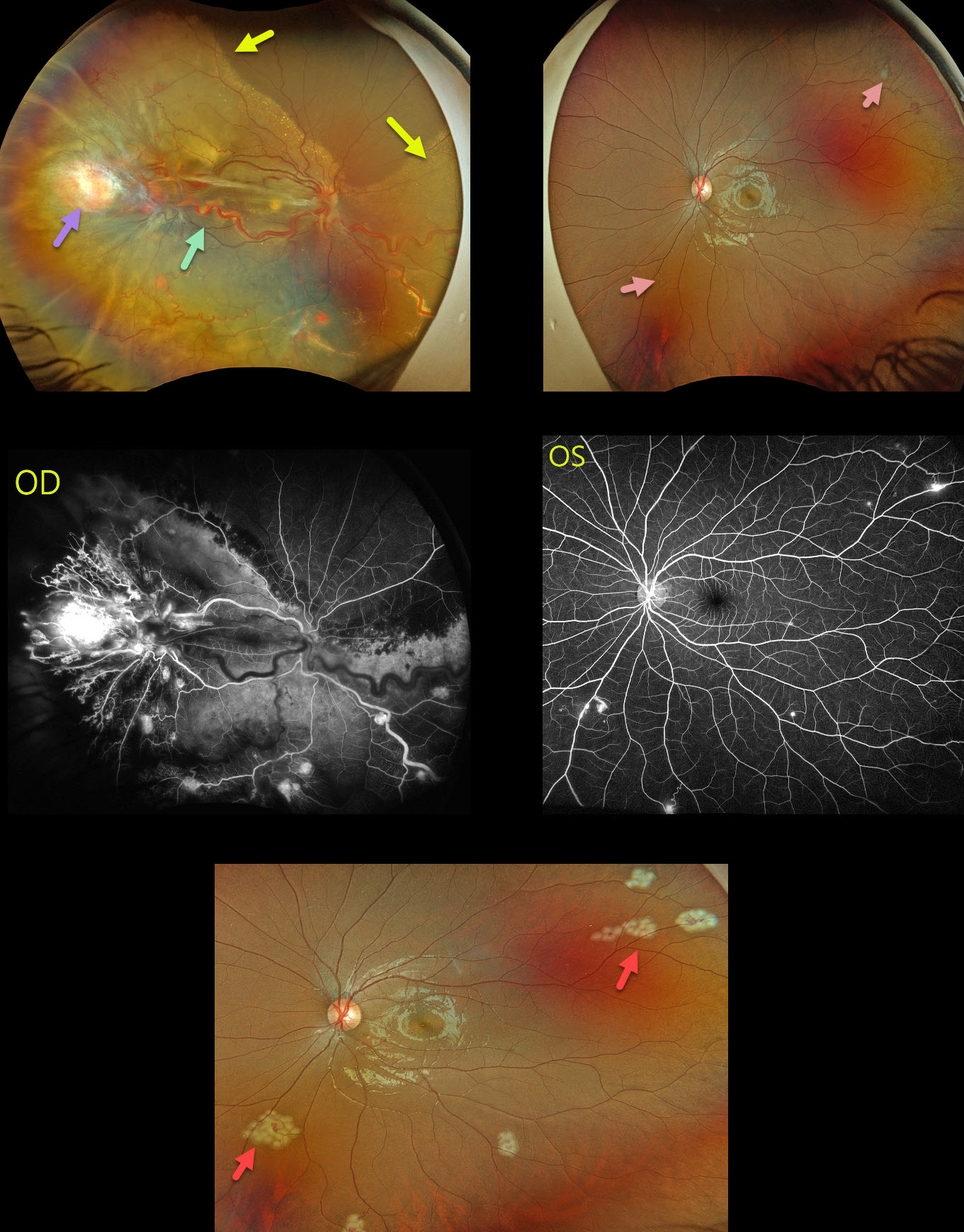
This 17YO female presented with a 3-month history of vision loss in her right eye. Vision was hand motion OD and 20/20 OS.
Optos color RGB imaging OD shows a large yellow-red angioma in the temporal periphery, associated with large dilated and draining vessels. There is another large angioma nasally, near the ora serrata, which is not well imaged. Multiple smaller angiomas are scattered throughout the temporal and inferior retina. A near-total exudative retinal detachment (RD) spares the superonasal retina. A few small peripheral angiomas are noted in her left eye. All tumors leaked angiographically.
The angiomas in the left eye were lasered during her initial examination. She then underwent lens-sparing vitrectomy in the right eye with membrane removal, endophotocoagulation, feeder vessel endodiathermy, and endoresection of the large temporal and nasal tumors. Mitomycin C was applied outside the posterior pole to reduce the risk of PVR, and silicone oil tamponade was used.
Our patient’s family history was negative. Genetic testing was subsequently positive for the von Hippel-Lindau (VHL) gene, which likely represents a de novo mutation.
Learning Points:
Retinal capillary hemangiomas are orange-red lesions associated with a prominent paired feeding and draining vessel. These benign vascular tumors need to be aggressively treated since they can cause severe vision loss or blindness from exudative RD, as in our patient. Patients require lifelong screening examinations for the development of new tumors even after successful treatment. Ultrawidefield imaging is helpful in detecting and monitoring these lesions.
Retinal capillary hemangioma is a predominant feature of VHL, an autosomal dominant condition caused by mutations in the VHL tumor suppressor gene. Patients develop benign and malignant tumors, including central nervous system hemangioblastoma, pheochromocytoma, and renal cell carcinoma.



