

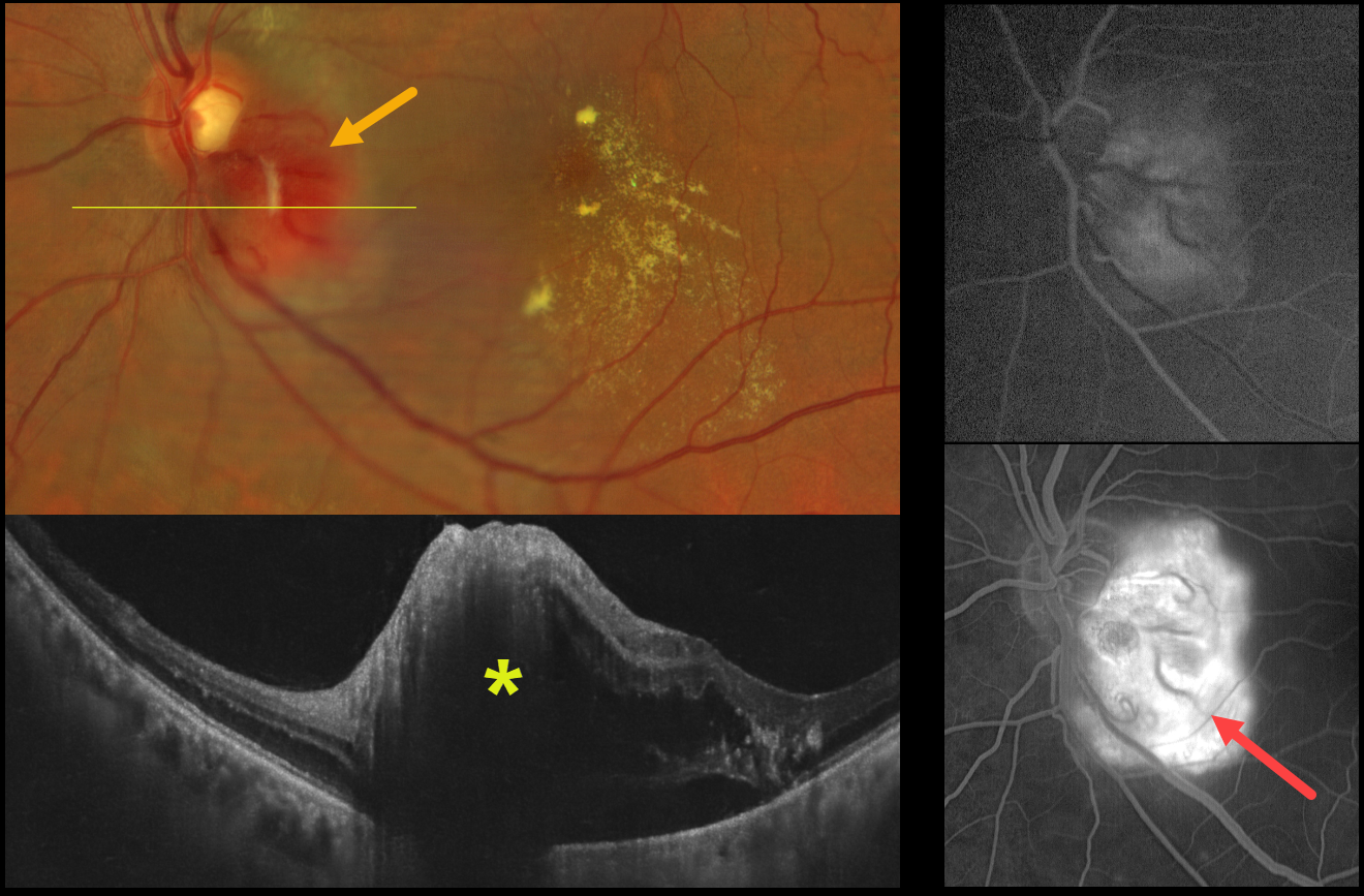
This healthy 51YO male presented with one month of vision loss in his left eye. Vision was 20/20 in his normal OD and 20/200 OS.
Color fundus photography shows an elevated orange-red juxtapapillary tumor along the inferotemporal edge of the optic nerve with associated macular lipid. OCT scanning reveals the lesion to be hyperreflective with associated outer retinal and subretinal fluid. The lesion leaked on fluorescein angiography.
Family history was negative for von Hippel–Lindau disease, and systemic evaluation, including brain MRI and renal ultrasonography, was unremarkable. Anti-VEGF therapy was recommended.
Learning Points:
Retinal capillary hemangiomas appear clinically as orange-red lesions, most commonly peripherally, and are associated with prominent, dilated, tortuous paired feeding and draining vessels. They can also appear on or around the optic nerve lesions, either on the surface (endophytic) as in our patient, or buried within the substance of the nerve (exophytic). Unlike peripheral lesions, juxtapapillary tumors often lack prominent feeder vessels. These lesions can cause severe vision loss or blindness from exudative retinal detachment and thus usually require aggressive treatment. Treatment options include thermal laser, photodynamic therapy, cryotherapy, and anti-VEGF injections.
More recently, oral belzutifan, an inhibitor of hypoxia-inducible factor 2 alpha, has emerged as a highly effective treatment for these lesions. In the LITESPARK-004, a Phase 2 trial, all 16 eyes improved without new lesions after a mean follow-up of 37 months (Wiley et al., Ophthalmology 2024;131;1324-1332).
Retinal capillary hemangioma is also a predominant feature of von Hippel-Lindau disease (VHL), an autosomal dominant condition caused by mutations in the VHL tumor suppressor gene. Patients develop benign and malignant tumors, including central nervous system hemangioblastoma, pheochromocytoma, and renal cell carcinoma. Rarely, sporadic, non-hereditary tumors may be found, as in our patient.



