

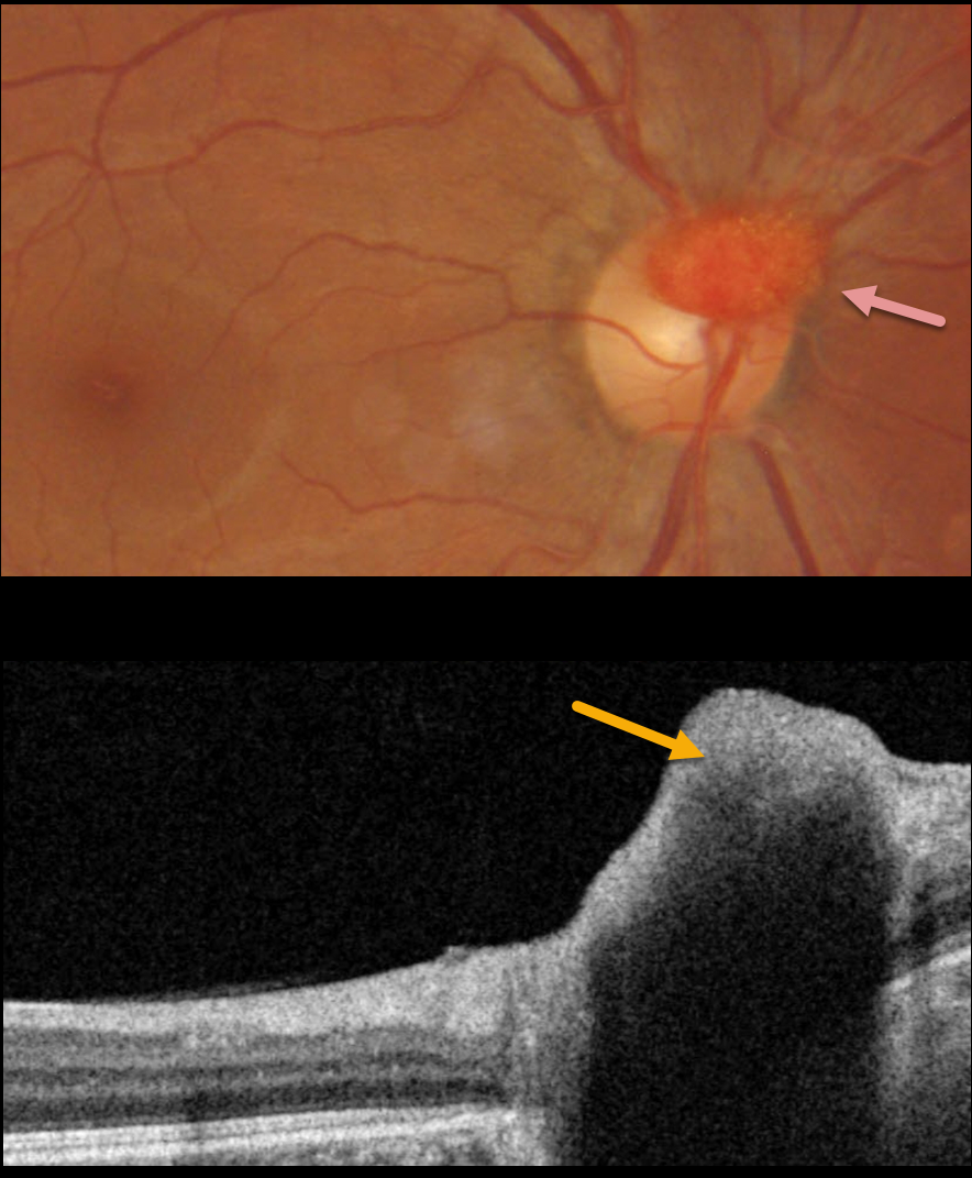
This 39YO healthy male was referred for an asymptomatic lesion in his right eye.
Fundus photography shows an elevated, vascularized lesion covering the superior optic nerve. The lesion was markedly hyperreflective on OCT.
On further questioning, there was a strong family history of Von Hippel-Lindau (VHL) disease, including his sister and maternal nephew. Genetic testing was ordered.
Learning Points:
Retinal capillary hemangioma is a predominant feature of VHL, an autosomal dominant condition caused by mutations in the VHL tumor suppressor gene. Patients develop benign and malignant tumors including central nervous system hemangioblastoma, pheochromocytoma and renal cell carcinoma.
Retinal capillary hemangiomas appear clinically as orange-red lesions, most commonly seen peripherally associated with a prominent paired feeding and draining vessel. They can also appear on or around the optic nerve lesions, either on the surface (endophytic) as in our patient, or buried within the substance of the nerve (exophytic).
These lesions can cause severe vision loss or blindness from exudative retinal detachment and thus usually require aggressive treatment. Treatment options include thermal laser, photodynamic therapy, cryotherapy, and anti-VEGF injections.
Since our patient is completely asymptomatic with no exudation, we will observe without treatment for now.



