

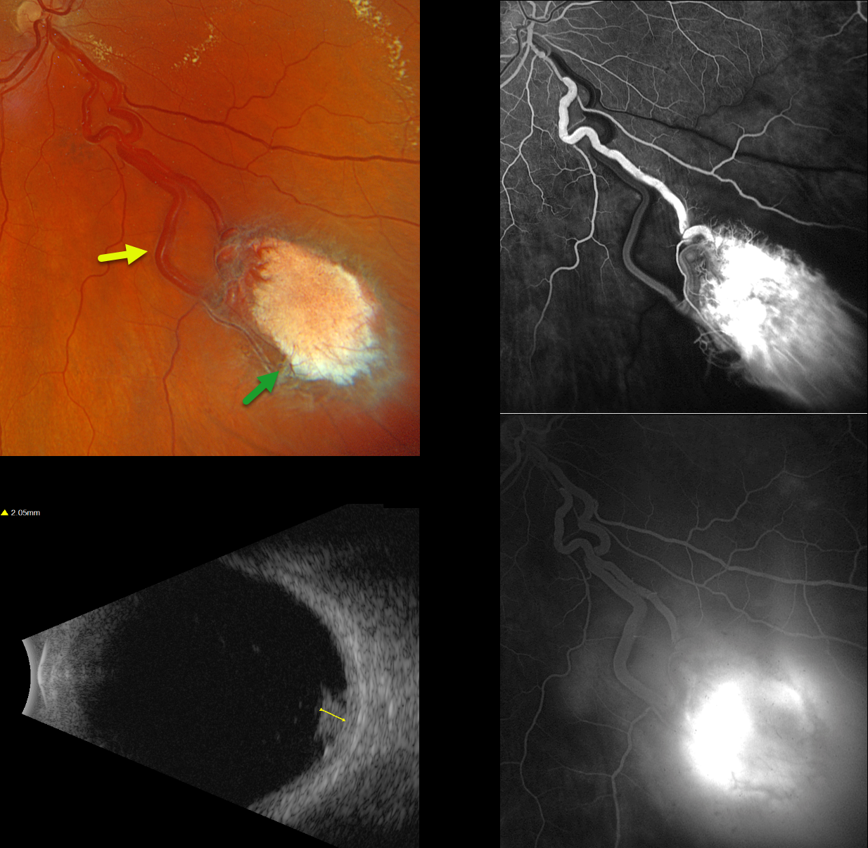
This healthy 42YO male was referred for an asymptomatic retinal lesion in his right eye. There was no family history of cancer or eye disease, although he had no knowledge about his birth father. Vision was 20/30 OD and 20/20 in his normal left eye.
Optos color RGB imaging shows an active endophytic retinal capillary hemangioma (RCH) in the inferonasal midperiphery. The tumor is supplied and drained by dilated, irregular vessels. The lesion shows profound leakage on fluorescein angiography. It measured about 2mm thick on B-scan ultrasonography. Genetic testing for the von Hippel-Lindau syndrome (VHL) gene was negative, as was MRI scanning of the brain, abdomen, and pelvis. Monthly intravitreal Avastin injections are planned to shrink the tumor, followed by thermal laser photocoagulation.
Learning Points:
Retinal capillary hemangiomas are orange-red lesions associated with a prominent paired feeding and draining vessel and can cause severe vision loss or blindness from exudative retinal detachment. These benign vascular tumors need to be aggressively treated. Common treatment options include thermal laser, cryotherapy, and anti-VEGF injections.
They are a predominant feature of VHL, an autosomal dominant condition caused by mutations in the VHL tumor suppressor gene. This prevents the degradation of hypoxia-inducible factor 1a (HIF-1a), which, in turn, causes the production of numerous growth factors, including VEGF (Kaelin, Drug Discoveries Today Disease Mechanisms 2005;2:225-231). Patients develop benign and malignant tumors, including central nervous system hemangioblastoma, pheochromocytoma, and renal cell carcinoma. Sporadic RCHs in the absence of the VHL syndrome are a rare occurrence (Singh et al, Ophthalmology 2001;108:1907-1911). The negative genetic testing fortunately ruled out VHL in our patient. Our patient’s lesion most likely resulted from a localized double-hit retinal VHL mutation.



