

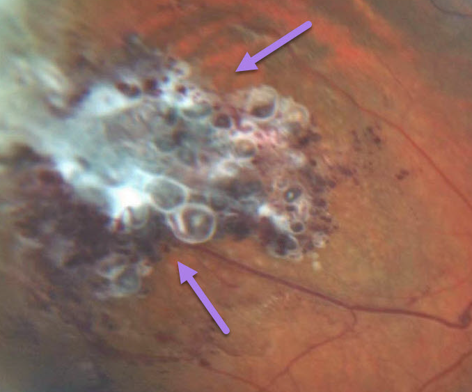
Retinal cavernous hemangiomas are usually unilateral, inner retinal grape-like clusters of aneurysms filled with dark venous blood. A gray fibrotic membrane often covers its superficial surface.
Vision is usually normal, and no treatment is required. Rarely, vision loss can occur from vitreous hemorrhage.
Learning Points:
Although most occur sporadically, some may be associated with an autosomal dominant neurocutaneous syndrome, including cutaneous and central nervous system hemangiomas. Neurologic imaging is therefore recommended.



