

This 32YO male presented with several weeks of decreased vision in the right eye. Vision was 20/200 OD and 20/20 in his normal OS. Examination revealed a dense vitreous hemorrhage, initially attributed to Valsalva retinopathy. He was immediately lost to follow-up and came back six years later with complaints of recent blurred vision with floaters. Vision was 20/50.
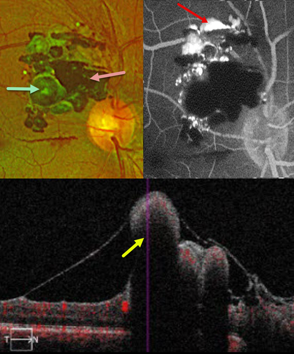
Optos color RG imaging shows a multilobulated mass superotemporal to the optic nerve, consisting of large saccular aneurysms with some overlying gliosis and preretinal blood. Fluorescein angiography shows superior layering of fluorescein within the vascular lesions, with blockage by blood. On OCT, the vascular abnormalities protrude into the vitreous with posterior shadowing.
Learning Points:
Retinal cavernous hemangiomas are rare, benign vascular lesions composed of thin-walled saccular venous aneurysms resembling clusters of grapes. Although most are asymptomatic and discovered incidentally, vision loss can occur from macular involvement or vitreous hemorrhage. Fluorescein angiography reflects the sluggish blood flow, classically showing plasma-erythrocyte layering as in our patient.
Although most are sporadic, some may be associated with an autosomal dominant neurocutaneous syndrome, including cutaneous and central nervous system hemangiomas. Neurologic imaging is therefore recommended, and our patient’s MRI results are pending.



