

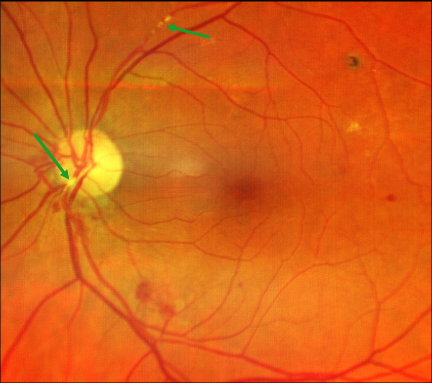
This 77YO male presented with unilateral left superior field loss in his left eye one day earlier. Vision was 18/20 bilaterally.
Color imaging shows retinal emboli along the proximal superotemporal arcade and overlying the inferonasal nerve heads. Some scattered retinal hemorrhages are also seen. The right fundus was normal. Visual field testing was normal.
He was immediately referred for a stroke evaluation, which was fortunately negative. Carotid Doppler showed bilateral 30% internal carotid artery stenosis. He was found to be in new-onset atrial fibrillation, which was the presumed source for the emboli, and was placed on oral anticoagulation therapy.
Learning Points:
Retinal emboli, which generally arise from either the heart or carotid arteries, can be calcific, platelet-fibrin, or cholesterol (Hollenhorst plaques). If large enough, they can obstruct a retinal arteriole and cause acute retinal ischemia, which is defined as transient monocular vision loss (TMVL, formerly known as amaurosis fugax), acute branch retinal artery occlusion, or acute central retinal artery occlusion.
TMVL requires an immediate referral to the nearest stroke center. Asymptomatic emboli can be more effectively evaluated for an embolic source with carotid Doppler and cardiac B-scan ultrasonography.



