

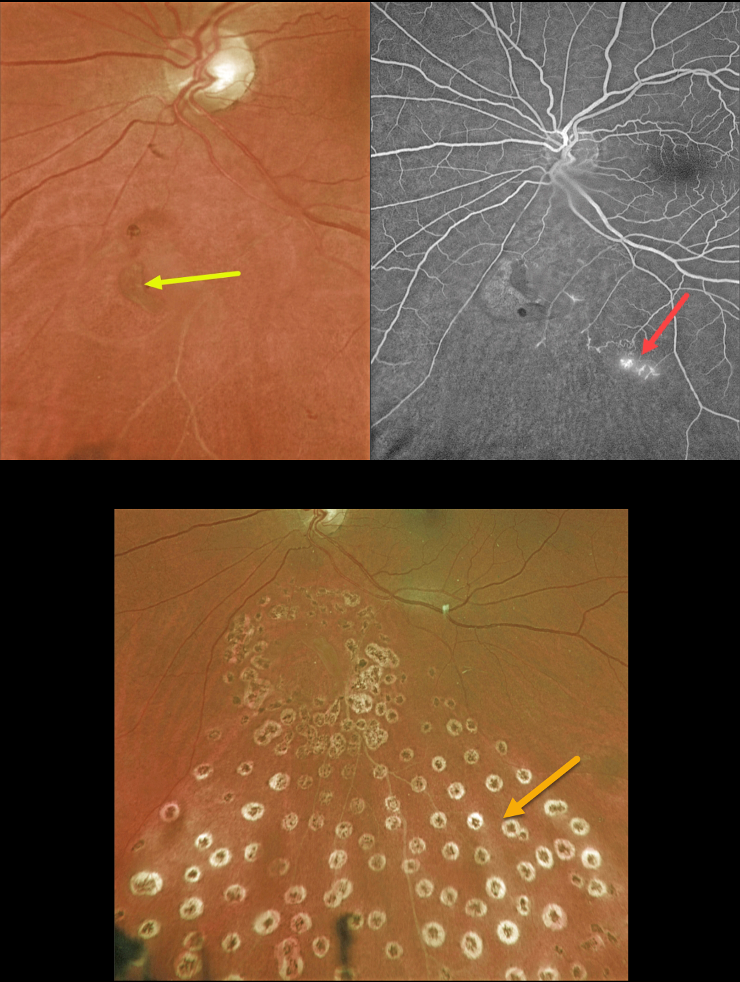
This 63YO female presented with symptomatic flashes and floaters in her left eye.
Optos color imaging shows a retinal tear just inferior to the nerve. An ischemic branch retinal vein occlusion (BRVO) is noted inferiorly. A posterior vitreous detachment (PVD) was noted clinically.
Fluorescein angiography confirmed the inferior ischemic BRVO with some small areas of retinal neovascularization. The tear appeared to be completely independent of the venous occlusion.
The retinal tear was surrounded by a few nearly confluent rows of laser photocoagulation burns, and inferior scatter laser was applied into the distribution of the BRVO. Fundus photography, a few months later, shows the laser scarring.
Learning Points:
Retinal tears are virtually always seen in the retinal periphery and are caused by an adherent vitreous pulling the retina anteriorly towards the vitreous base, most commonly during an acute PVD.
Our patient’s tear is unusual due to its quite posterior location and the flap occurring along its posterior edge. This implies that the vitreous was tugging towards the optic nerve. The optic nerve is usually the last site for the vitreous to release from during an acute PVD (Johnson, AJO 2010;149:371-382).
We hypothesize that the vitreous was completely detached except at the optic nerve and just inferiorly, and the tear was created from anterior to posterior traction when the vitreous separated from the nerve.



