

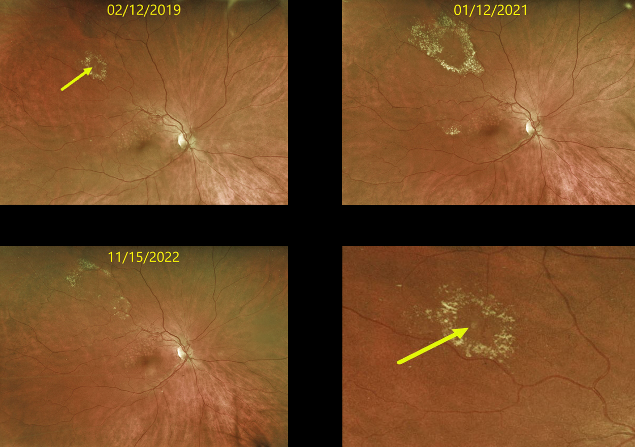
This 63 YO female originally presented in 2019 with a history of a major superotemporal branch retinal vein occlusion (BRVO). Successful macular laser was performed for macular edema.
When initially examined, a midperipheral retinal venous macroaneurysm (RVM) with some surrounding lipid was noted. This RVM has been observed due to its distant location from the fovea. The series of photos over several years shows the exudation to wax and wane. At the last examination on 11/15/22, there was almost complete lipid resolution. Vision remains at 20/25.
Learning Points:
In 1990 Cousins et al reported that RVMs can be an additional finding in occlusive venous disease, always in association with collateral vessels or capillary or arterial macroaneurysms (AJO 1990;109;567-1700).
These vascular abnormalities tend to have a higher rate of intraretinal lipid (40%) but are less likely to affect central vision since they are usually outside the macula.
They tend to have a higher association with retinal ischemia, thus being at a higher rate of developing retinal neovascularization (32%). The lesions will usually involute, as was the case in our patient.



