

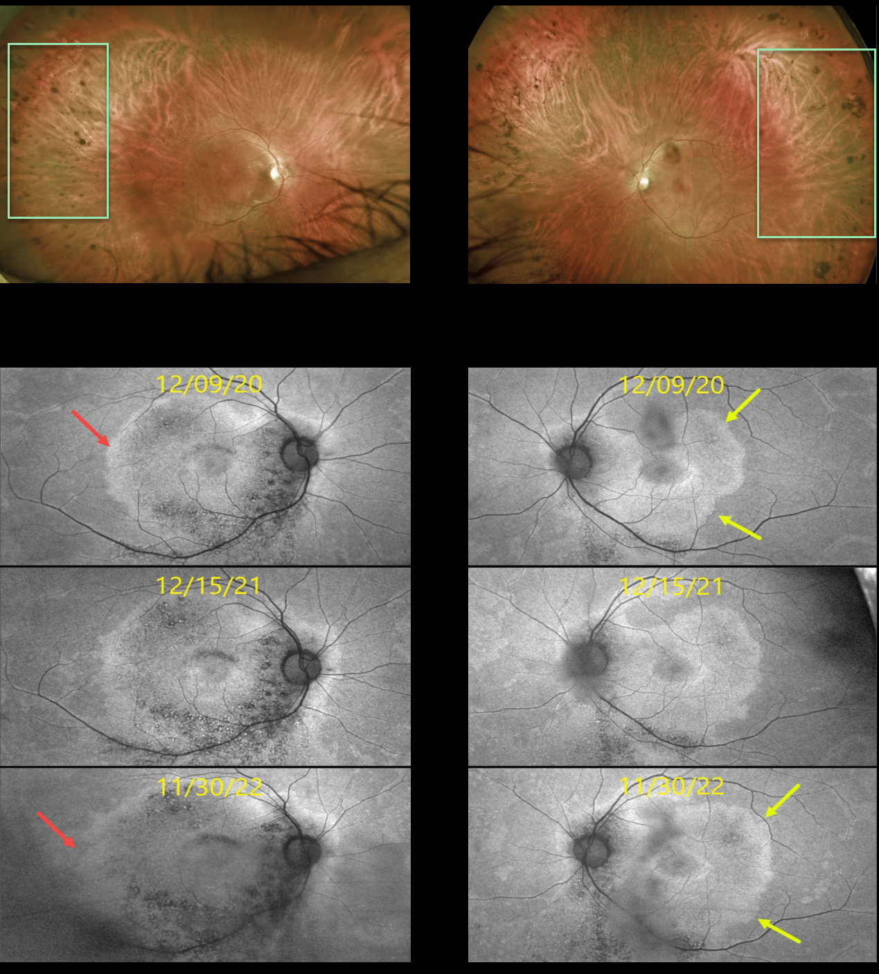
This 53YO male presented on 12/9/20 with a history of known retinitis pigmentosa (RP). He noticed bilateral blurred vision that had been worsening for about 6 months. Vision was 20/50 OD and 20/30 OS.
Optos color imaging shows fairly normal appearing maculas with a fairly-well circumscribed border of pigmentary loss and migration extending anteriorly from the mid-peripheries.
However, fundus autofluorescence (FAF) reveals subclinical findings of increased macular hyper-FAF sparing each fovea, with hypo-FAF more inferiorly. Subsequent genetic testing revealed a pathogenic RP1 mutation.
FAF shows gradual progressive changes over the 2 years we have been following him. The temporal margins of the hyper-FAF in his right and left eyes have significantly enlarged. This is due to the unmasking of the underlying RPE with progressive photoreceptor atrophy.
Learning Points:
FAF visualizes fluorophores, endogenous compounds that spontaneously fluoresce without the need for an external dye. The main fundus fluorophore is lipofuscin, which resides within the RPE lysosomes.
The photoreceptor outer segments absorb some of the autofluorescent excitatory light, and thus normally diminish the FAF signal.
In RP, loss of the outer segments unmasks this signal, leading to increased FAF. Many patients with RP will show a hyper-FAF ring between clinically normal and abnormal retina, which likely represents retina ‘at risk’, showing the junction of functional and dysfunctional retina.
See Schmitz-Valckenberg et al (Progress Retinal Eye Research 2021;81:100893) for an excellent general review of FAF.
For further reading on monitoring RP with FAF, see Robson et al (Retina 2011;31:1670-1679) and Lee et al (Ophthalmology Retina 2018;2;1062-1070).



