

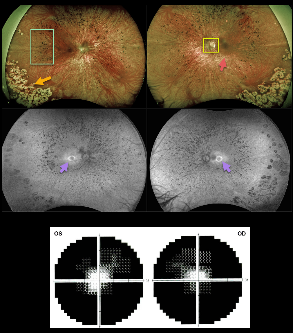
This 35YO female has classic findings for retinitis pigmentosa. Optos imaging shows somewhat pale nerves, narrowed retinal vessels, and peripheral intraretinal pigment migration (bone spicules). There are also confluent cobblestone-like changes in the inferotemporal peripheries.
Fundus autofluorescence (FAF) shows a hyper-FAF ring around the central macula, indicating preserved photoreceptors that may be at-risk for future degeneration.
Visual fields are markedly constricted bilaterally.
Learning Points:
Photoreceptors normally inhibit RPE cells, so any process involving photoreceptor loss can cause intraretinal RPE migration that mimics retinitis pigmentosa, including chronic retinal detachment and blunt trauma.



