

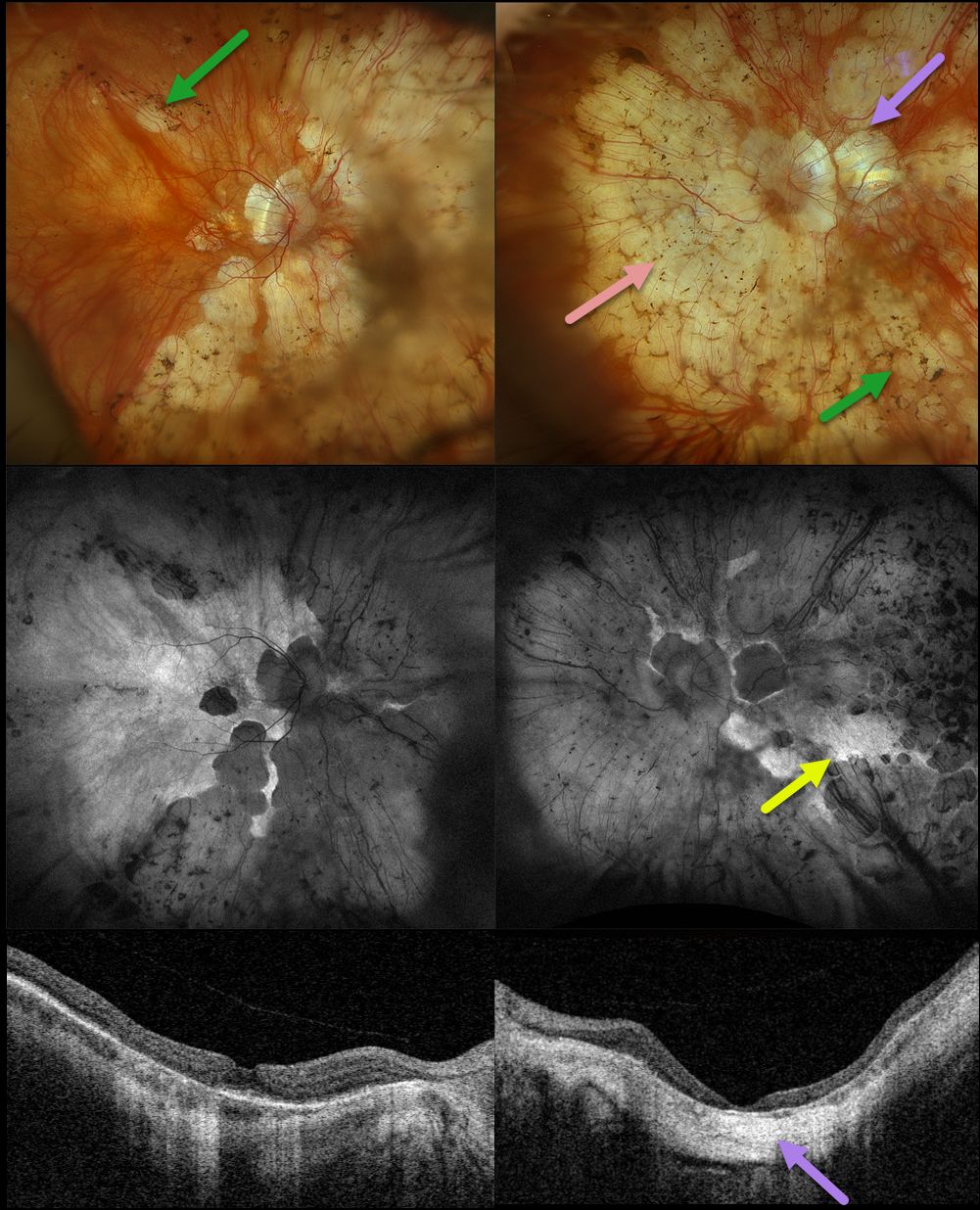
This 78YO female has been followed by us for several years for retinitis pigmentosa (RP) and degenerative myopia. Vision is stable at 20/30 OD and hand motion OS. Multimodal imaging shows a fascinating fusion of findings for these two disorders.
Optos color RGB imaging shows typical RP findings of bone spicules (intraretinal pigment migration) extending from outside the arcades into the peripheries, superimposed on severe degenerative myopic changes.
Fundus autofluorescence (FAF) shows hypo-FAF within the areas of myopic atrophy, with hyper-FAF more peripherally from the RP-induced outer retinal atrophy.
A macular staphyloma is also seen in the left eye and confirmed on swept-source OCT. The degenerative myopic changes just spare the right macular center, which has no staphyloma.
Learning Points:
Genetic testing showed pathogenic heterozygous mutations for OCA2 and RPGR. Fortunately, the RP spares each macula. Her myopic degeneration has been stable, and we are hopeful that her central vision will be preserved in her right eye.



