

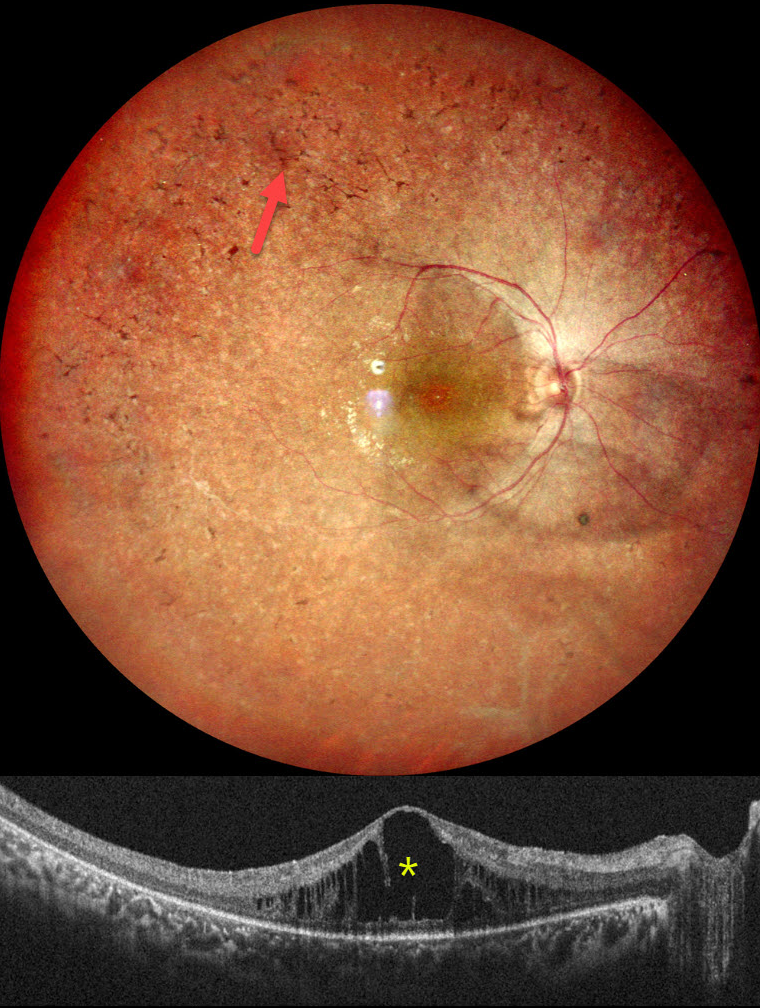
This healthy 18YO female complained of night blindness for a few years. Her mother and sister have a history of retinitis pigmentosa (RP). Vision was 20/90 OU.
Pseudocolor SLO imaging of her right eye shows typical RP scarring with intraretinal pigment migration extending from the macula into the retinal periphery. There is loss of the foveal reflex and cystoid macular edema (CME). Identical findings were noted in the OS (not shown).
Learning Points:
RP is a clinically and genetically heterogeneous group of inherited retinal disorders, which can present sporadically or with any inheritance pattern (autosomal dominant, autosomal recessive, sex-linked, or mitochondrial). It is characterized by diffuse, progressive dysfunction of predominantly rod photoreceptors, followed by RPE degeneration with intraretinal pigment migration. Visual impairment usually presents as night blindness and progressive visual field loss.
CME is observed in up to 20% of RP patients, often without angiographic leakage. Pathological mechanisms include vitreous traction/epiretinal membranes or a breakdown of the blood-retinal barrier due to inflammation or RPE/Muller cell dysfunction (Gaudric et al, Progress in Retinal and Eye Research 2022;91:101092). First-line treatment is topical or oral carbonic anhydrase inhibitors (CAIs). Other options for refractory CME include steroids (topical, periocular, or intravitreal), anti-VEGF injections, or vitrectomy for a tractional component. Our patient was given topical CAIs but was immediately lost to follow-up.



